Case of April 2022
For completion of the online quiz, please visit the HKAM iCMECPD website: http://www.icmecpd.hk/
Clinical History:
A 40-year-old man, chronic hepatitis B carrier, was admitted for right upper quadrant abdominal pain and fever. Laboratory test revealed mild leukocytosis (WCC up to 15 x 109/L) which subsequently normalized after starting intravenous augmentin. Liver, renal function tests remained normal. He underwent ultrasound, contrast CT abdomen during hospitalization. Tumour markers with AFP, CA 19.9 and CEA levels were normal. His abdominal pain and fever subsided with medical treatment and was fit for discharge afterwards. He underwent PET CT one week later.
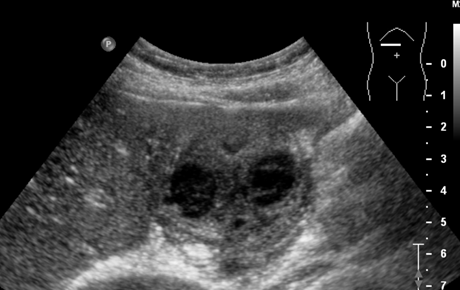
Initial abdominal ultrasound (TS)
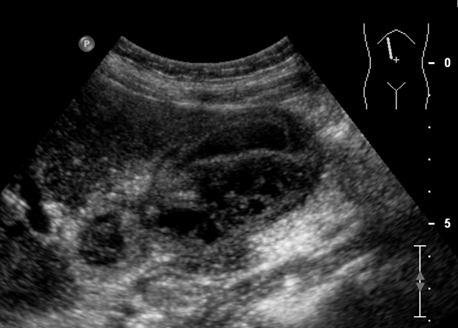
Ultrasound (LS)
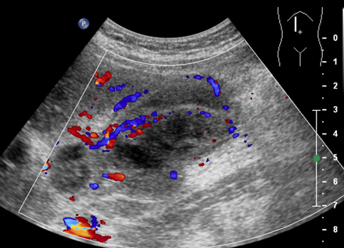
USG LS (with colour doppler)
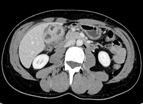
Contrast CT abdomen (axial)
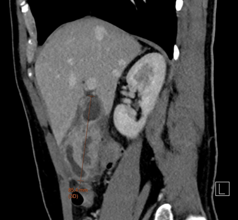
Contrast CT abdomen (sagittal)

PET CT fusion images (axial) 1 week after
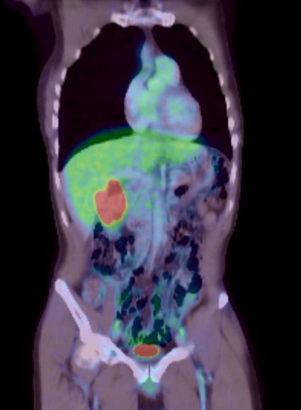
(PET CT fusion coronal)
![]()