Case of January 2020
For completion of the online quiz, please visit the HKAM iCMECPD website: http://www.icmecpd.hk/
Clinical History:
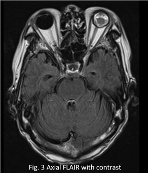
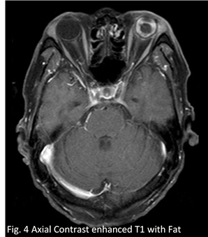
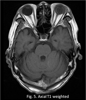
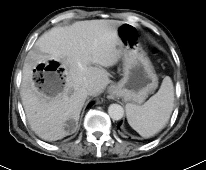
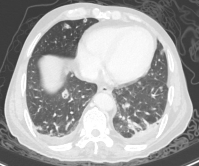
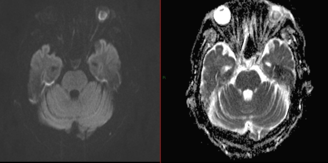
56-year-old male patient was admitted for abdominal pain and fever. Patient was an alcoholic and had history of poorly controlled DM. Laboratory findings showed elevated white cell count and inflammatory markers. Contrast CT Abdomen was performed as initial investigation. Later the patient developed confusion and contrast MRI of the brain was performed for assessment.
|
Fig. 1 Contrast CT Abdomen |
Fig. 2 Contrast CT in lung window |
 |
 |
 |
|
|
Fig. 6 Diffusion weight imaging (DWI) (left side) and ADC map (right side) of our patient, demonstrating restricted diffusion of the thickened uvea. |
|
![]()