Case of January 2016
For completion of the online quiz, please visit the HKAM iCMECPD website: http://www.icmecpd.hk/
Clinical History:
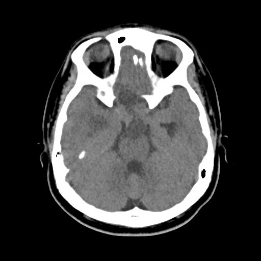
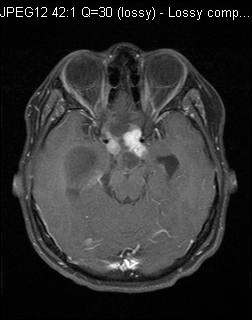
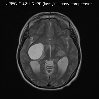
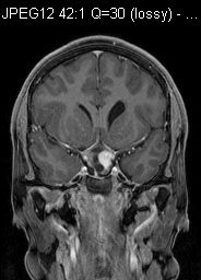
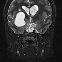
A 13-year-old boy has history of poor vision since 3 years old. Otherwise, no focal neurological sign or abnormal growth was noted. CT and MR of brain were performed to investigate for visual loss.
Fig 1. Plain CT Fig 2. MR-T1 weighted axial, with contrast
Fig 3. MR-T2 weighted axial Fig 4. MR-T1 weight coronal, with contrast
Fig 5. MR-T2-weighted coronal
