Case of April 2016
For completion of the online quiz, please visit the HKAM iCMECPD website: http://www.icmecpd.hk/
Clinical History:
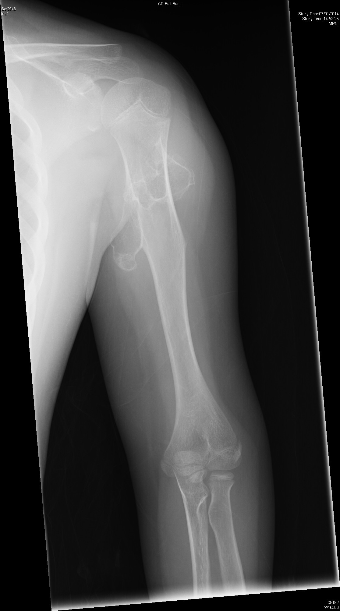
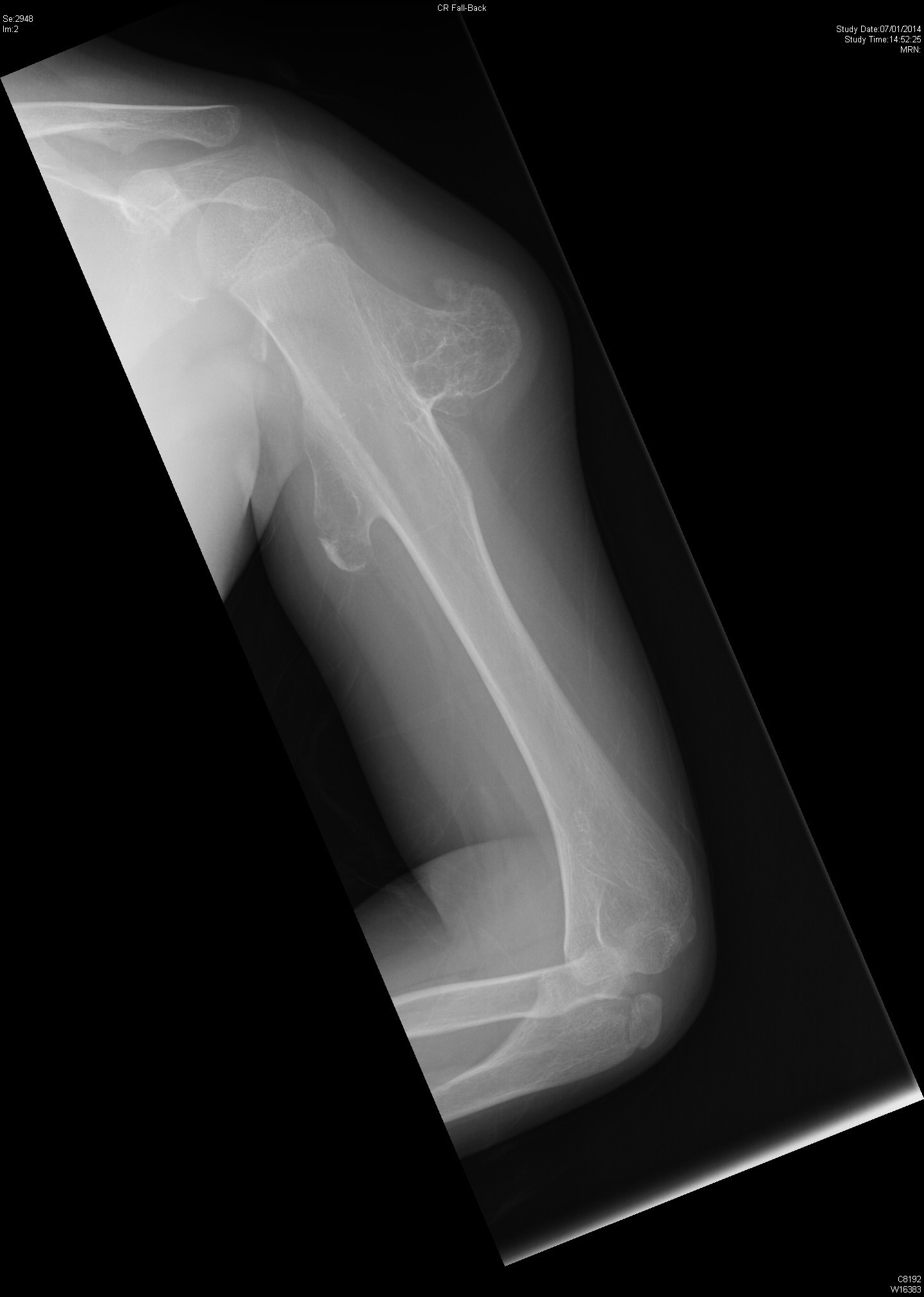
A 13 years old boy with good past health presented as left shoulder mass for several years without any pain. The mass was gradually increasing in size. He has strong family history of bony masses. His father had history of bony mass excision in his teenage. Physical examination found 4cm diameter bony mass on the lateral aspect of left upper arm. Range of movement of his left shoulder was full.
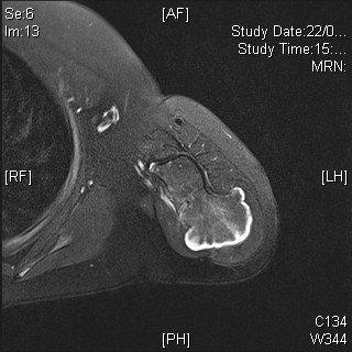
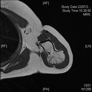
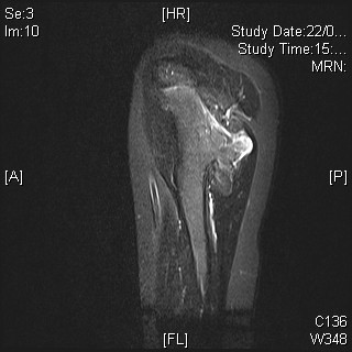
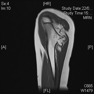
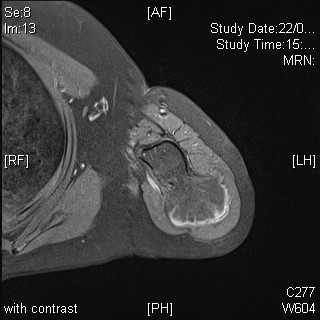
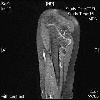
Work up includes plain radiographs of his left humerus (Picture 1-2) and subsequent MRI (Picture 3-8).
Picture 1
Picture 2
Picture 3 (Axial T2 with fat saturation)
Picture 4 (Axial T1)
Picture 5 (Sagittal STIR)
Picture 6 (Sagittal T1)
Picture 7 (Axial T1 post-gadolinium)
Picture 8 (Sagittal T1 post-gadolinium)
