Case of September 2015
For completion of the online quiz, please visit the HKAM iCMECPD website: http://www.icmecpd.hk/
Clinical History:
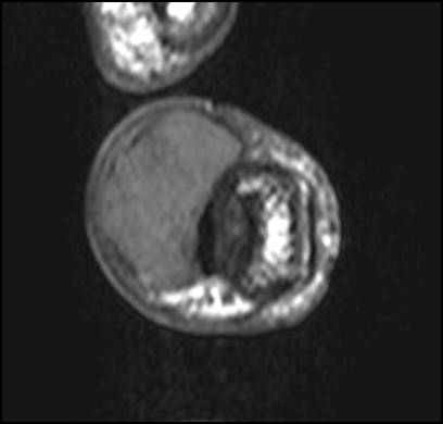
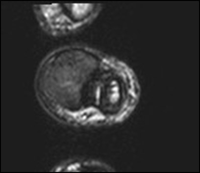
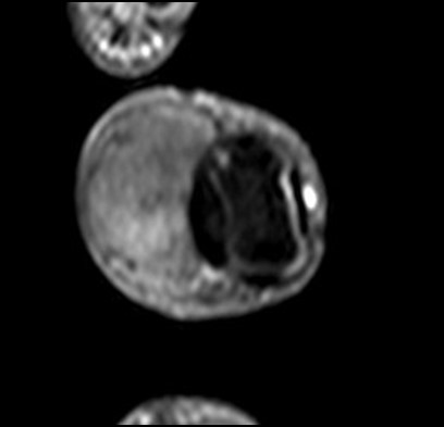
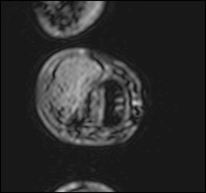
A 66 year-old gentleman presented with left middle finger swelling for 2 years. It was slowly increasing in size, with no pain or numbness. Physical examination showed a firm multi-lobulated mass of about 1.5cm over the volar aspect of left middle finger at the distal interphalangeal joint level. Magnetic resonance imaging (MRI) was performed (4 images – upper row: axial T1-weighted image, axial T2-weighted image; lower row: post-contrast fat-suppressed axial T1-weighted image, axial T2-weighted gradient-echo image)
MRI


