Answer of June 2015
For completion of the online quiz, please visit the HKAM iCMECPD website: http://www.icmecpd.hk/
Clinical History:
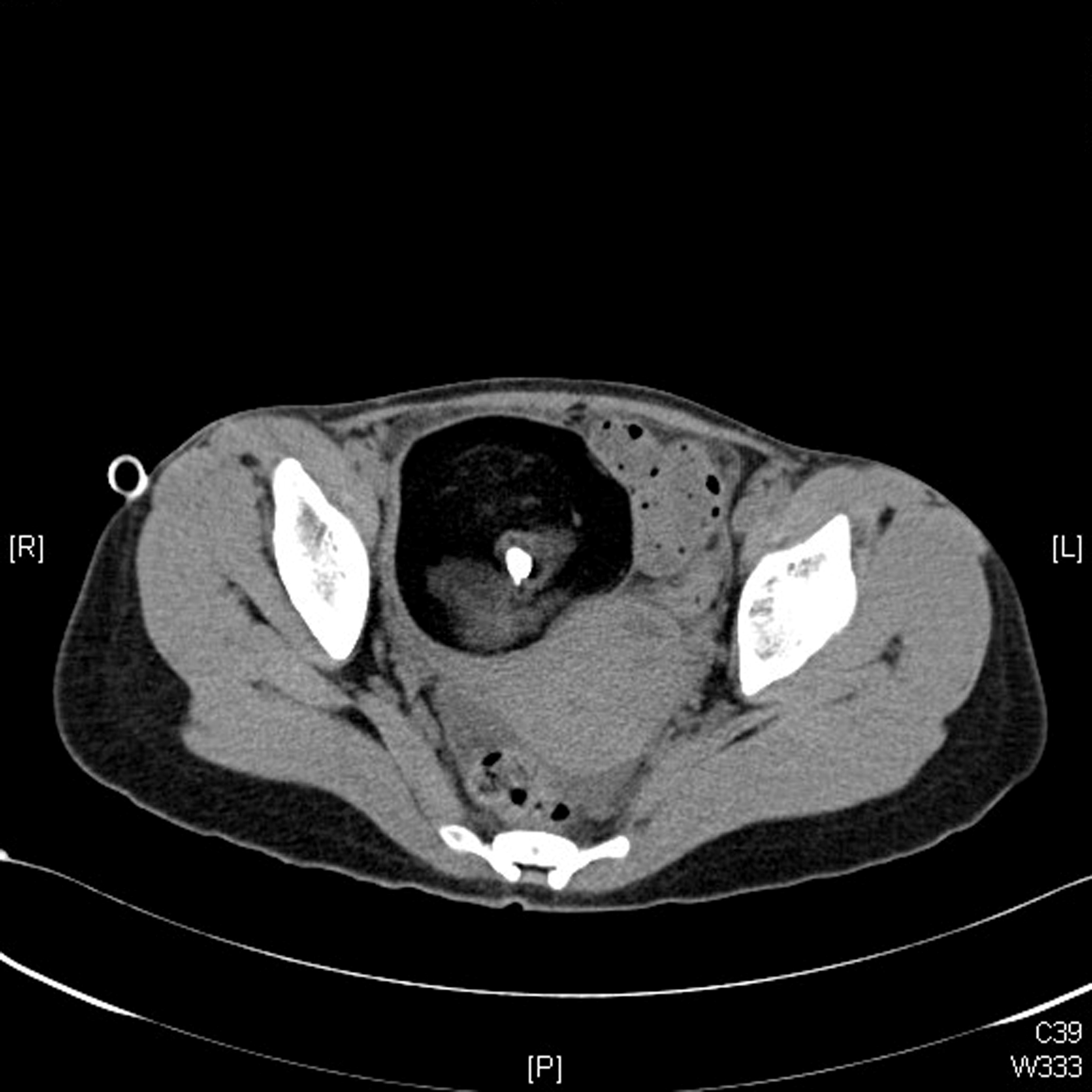
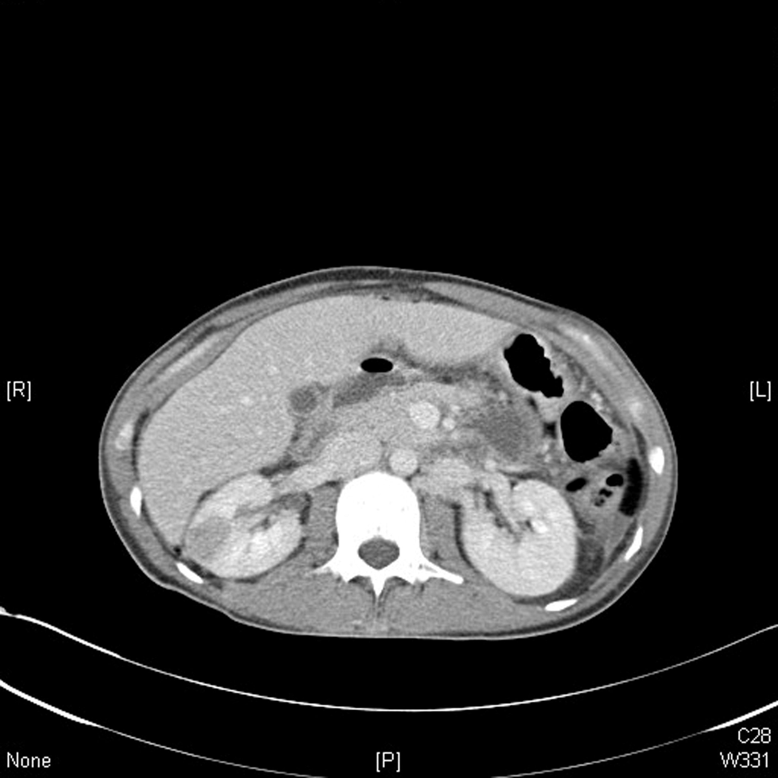
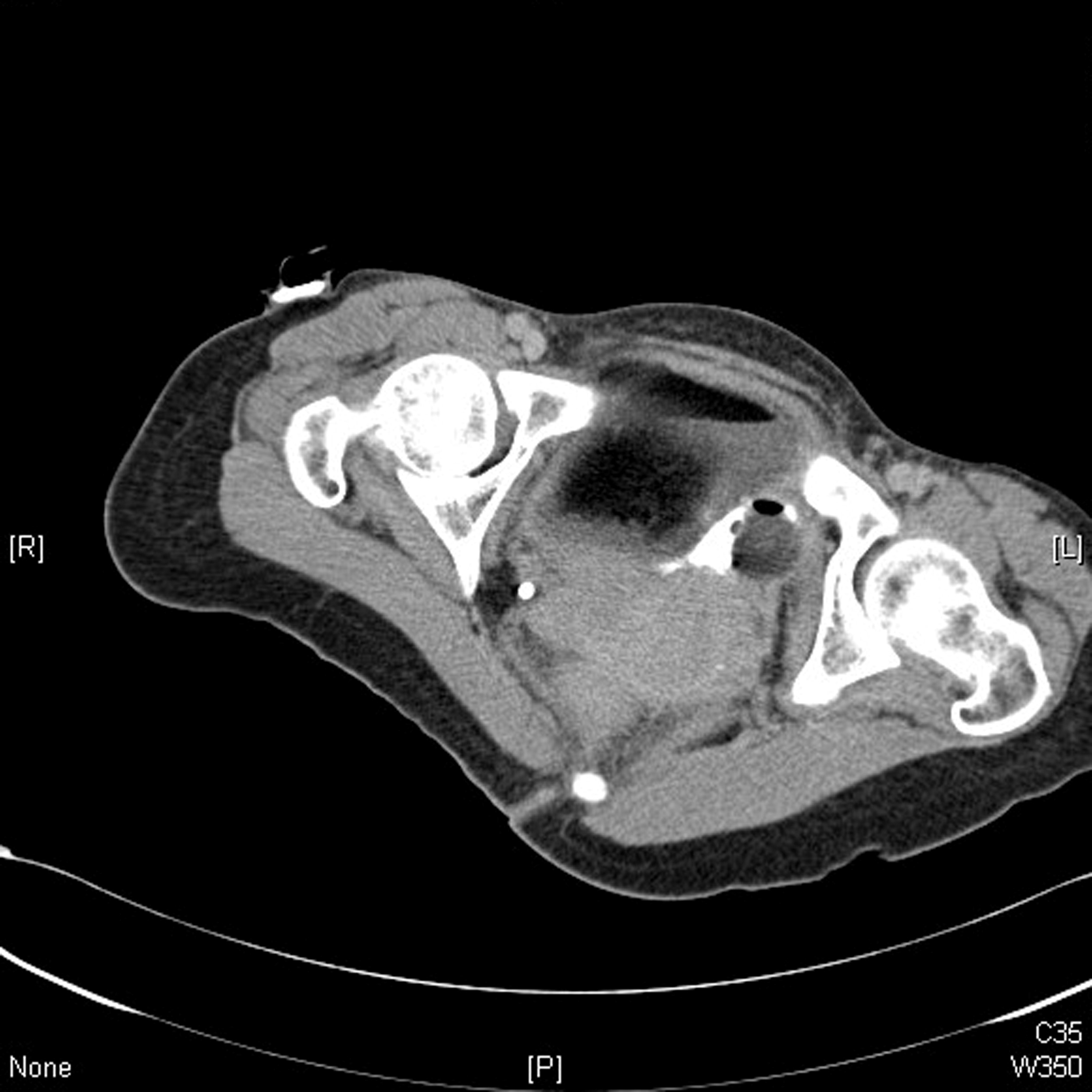
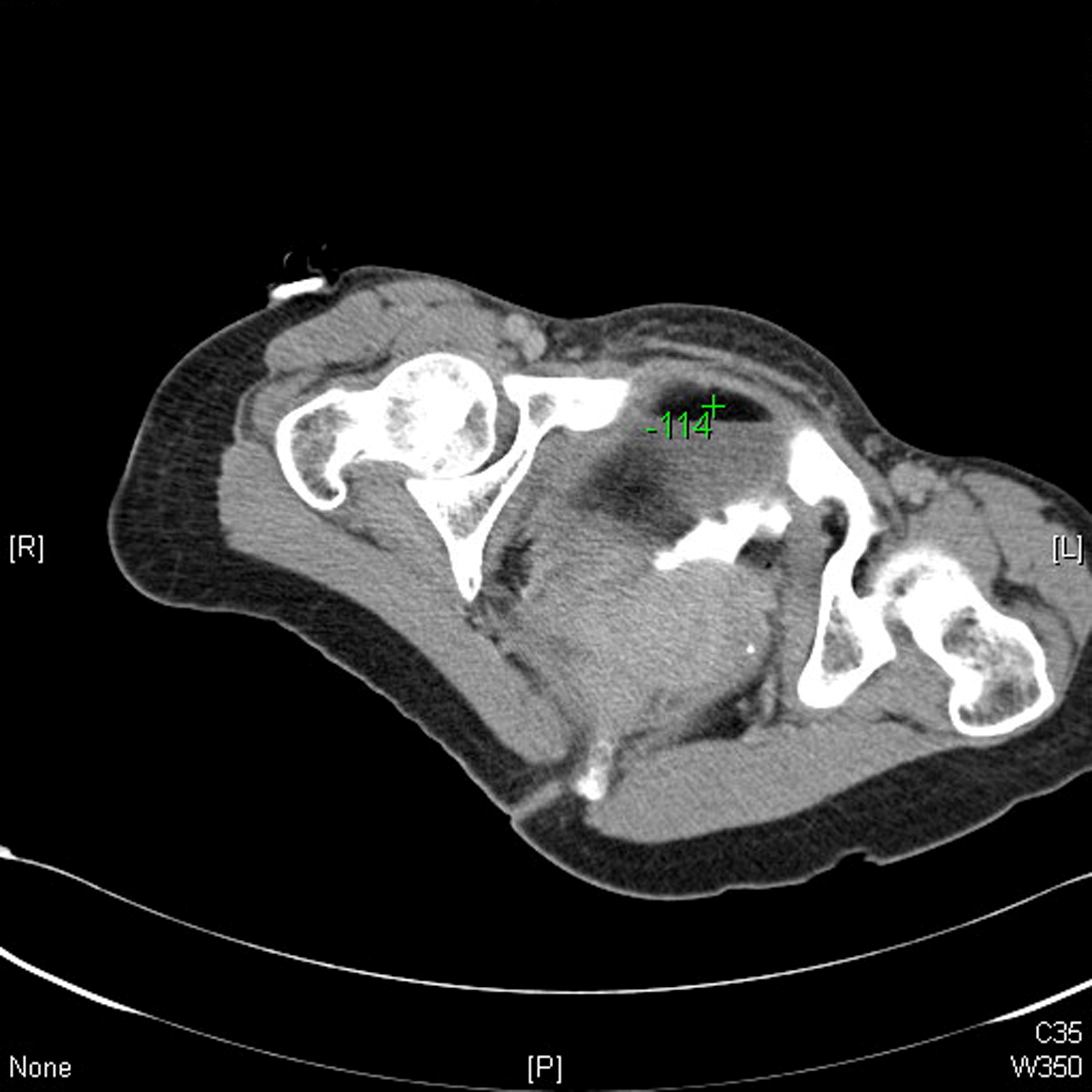
A 40-year-old lady presented acutely with diffuse left sided abdominal pain for 1 day and fever for 1 week. She was febrile on arrival with tenderness and guarding in the left iliac fossa. Haemoglobin was 10.5g/dl and white cell count was 14.9. Urgent CT scan (Non-contrast and contrast scans) was performed assessment (Fig. 1-4):
Fig 1
Fig 2
Fig 3
Fig 4
Diagnosis:
Discussion:
Ovarian cystic teratoma accounts for 5-25% of all ovarian masses. It can present bilaterally in 8-15% of cases. Most are asymptomatic but symptoms can include pain and mass symptoms. Complications can include torsion (16%), malignant degeneration (2%) and rupture (1-2%).
Spontaneous rupture is uncommon and can result from undetected torsion, trauma, or pressure from gravid uterus. Fatty sebaceous material can leak into the intraperitoneal space causing chemical peritonitis. This would result in a presentation of acute pain and abdominal distention.


