Answer of May 2024
For completion of the online quiz, please visit the eHKAM LMS website.
Clinical History:
A 50-year-old gentleman presented with fever, sore throat, dysphagia, and recent unintentional weight loss. Flexible laryngoscopy noted a large friable nasopharyngeal mass, and cervical lymphadenopathy was palpable. Contrast-enhanced computed tomography of the neck was performed.
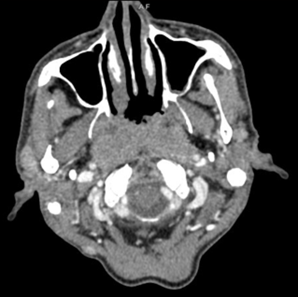
Figure 1. Axial contrast-enhanced computed tomography (CT) at the level of nasopharynx
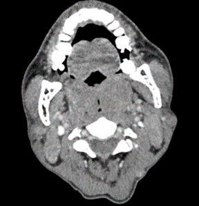
Figure 2. Axial contrast-enhanced CT at the level of oropharynx
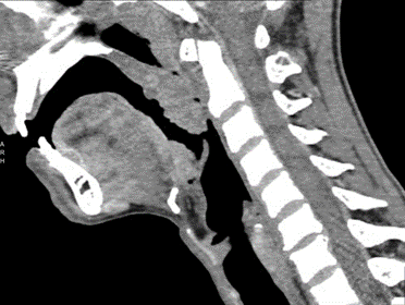
Figure 3. Sagittal-contrast-enhanced CT at the midline
DIAGNOSIS
Nasopharyngeal lymphoma
IMAGING FINDINGS
Figure 1 demonstrates a homogeneous midline nasopharyngeal mass, and bilateral enhancing intraparotid lymphadenopathy. Figure 2 shows associated enlargement of the palatine tonsils (‘kissing’), likely predisposing to airway compromise. Significantly enlarged bilateral level II cervical lymph nodes also noted. Figure 3 shows enlargement of the Waldeyer’s ring; including pharyngeal, palatine, and lingual tonsils. No gross bone erosion is seen in the images.
DISCUSSION
The diagnosis in this case is lymphoma of the nasopharynx, which was confirmed with bilateral tonsillectomy showing peripheral T-cell lymphoma with T-follicular helper cells differentiation. An initial nasopharyngeal biopsy had shown no malignant cells.
This patient developed stridor after the CT examination, and was prophylactically intubated.
Lymphoma of the nasopharynx is the second most common nasopharyngeal malignancy behind nasopharyngeal carcinoma, though accounting for less than 10% of nasopharyngeal malignancies. It is commonly due to non-Hodgkin lymphoma (as in the case of this gentleman), particularly diffuse large B cell lymphoma, while Hodgkin lymphoma is less common at this location.
When presented with a mass at the nasopharynx, several imaging features increase suspicion for the diagnosis of lymphoma. Involvement of the lymphoid tissue at the Waldeyer’s ring is classical, comprising the pharyngeal, tubal, palatine, and lingual tonsils. Involvement of the intraparotid and submandibular lymph nodes is a feature more commonly seen in lymphoma compared to its major differential diagnosis of nasopharyngeal carcinoma (NPC). Other supporting features include midline nasopharyngeal location (in contrast to involvement of the lateral nasopharynx and fossa of Rosenmuller in NPC), lack of necrosis, and absence of invasion to parapharyngeal spaces and absence of skull base erosion, both of which are involved early in NPC but less commonly involved by lymphoma.


