Answer of September 2023
For completion of the online quiz, please visit the eHKAM LMS website.
Clinical History:
A 27-year-old lady complains of right leg swelling.
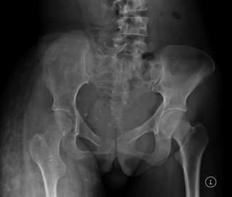
XR Pelvis
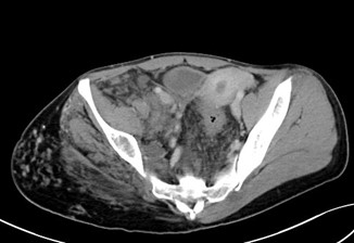
Contrast CT Pelvis
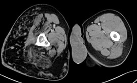
Contrast CT Upper Thighs
DIAGNOSIS
Klippel-Trénaunay syndrome
IMAGING FINDINGS
XR Pelvis - The radiograph shows multiple calcified pelvic phleboliths and hypertrophic changes over the right-side raising suspicion of local gigantism. The relative hypoplasia of the right hemipelvis and right femur is suggestive of underlying chronic or perhaps developmental processes.
Contrast CT - The contrast-enhanced CT demonstrates right gluteal muscle atrophy. right hemipelvis hypoplasia, right lower limb hypertrophy, presence of venous malformation and pelvic phleboliths.
Klippel-Trénaunay syndrome classically comprises a triad of:
● cutaneous capillary malformations: port wine nevi
● limb overgrowth: bony or soft tissue hypertrophy of an extremity (localized gigantism)
● varicose veins or venous malformations of unusual distribution
The diagnosis of Klippel-Trénaunay syndrome is usually made when any two of the three features are present. Patients usually present in infancy. Features are often unilateral and typically affect one limb; capillary malformations may be absent in the atypical form. It may be diagnosed in utero.
Hypertrophy
Enlargement of the extremity consists of bone elongation, circumferential soft-tissue hypertrophy or both. This often manifests as leg-length discrepancy, although any limb may be affected.
Capillary malformations
This is the most common cutaneous manifestation of Klippel-Trénaunay syndrome. Typically, capillary malformations involve the enlarged limb, although skin changes may be seen on any part of the body. The lower limb is the affected site in ~95% of patients.
Plain radiograph
On conventional radiography, bone elongation contributing to leg length discrepancy, soft-tissue thickening, or calcified phleboliths may be seen.