Answer of October 2022
For completion of the online quiz, please visit the HKAM iCMECPD website: http://www.icmecpd.hk/
Clinical History:
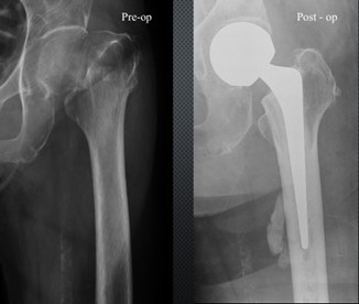
A 50 year-old lady suffered from a left femoral neck fracture after falling at home. She underwent left hip arthroplasty.
Preoperative and postoperative XRs
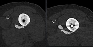
Postoperative CT
DISCUSSION
Cement arteriovenogram refers to the leakage of cement through nutrient vessels in the bone. It is commonly present as a peri-diaphyseal radio-opacity.
Schiessel et al. reported the presence of nutrient canal vessel in 26.4% radiographs of preoperative and postoperative radiographs of hip replacement and also reported its anatomical location about 170 ± 25 mm distal to greater trochanter.
CT is recommended on detection of a radio-opaque density on postoperative radiographs to differentiate an extraosseous breach from an intra-vascular extrusion of cement. Findings in three cases suggest that cement retrograde extrusion into nutrient vessels following hemiarthroplasty is a benign complication of modern cementing techniques involving pressurization. The site of cement extrusion into the nutrient foramina displays a constant topography. A CT scan of the femur can be done on detection of a radio-opaque density on postoperative radiographs to differentiate an extraosseous breach from an intra-vascular extrusion of cement.
Panousis et al. reported PMMA within a small diameter vessel with no valvular constrictions resulting in retrograde arteriogram of the nutrient artery of the femur which coupled with the extent of intraoperative endosteal bleeding. The plain radiographs confirmed linear extrusion of PMMA along the course of nutrient artery proximally and medially with no valvular constrictions resulting in the retrograde arteriogram of the nutrient artery of the femur.

