Answer of June 2022
For completion of the online quiz, please visit the HKAM iCMECPD website: http://www.icmecpd.hk/
Clinical History:
A 35-year-old lady (gravida 1, para 0) suffered from severe primary postpartum hemorrhage due to uterine atony after vaginal delivery, which was subsequently controlled by Barki Balloon and B-lynch compression suture. B-lynch suture is a widely adopted surgical technique for postpartum hemorrhage by placement of compression sutures onto the uterine body with an intent to control the bleeding. Contrast enhanced CT abdomen (Figure, A and B) was performed 15 days after delivery for persistent fever.
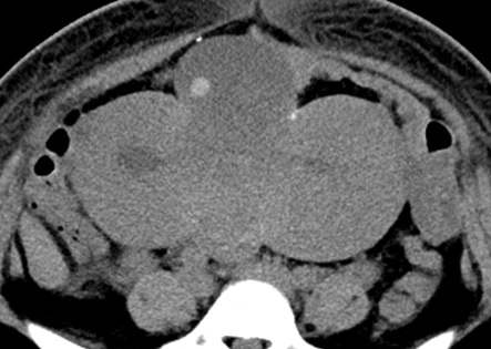
CT Abdomen plain– Axial
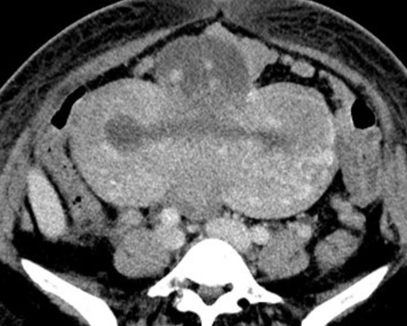
CT Abdomen with contrast – Axial
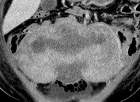
CT Abdomen with contrast – Coronal
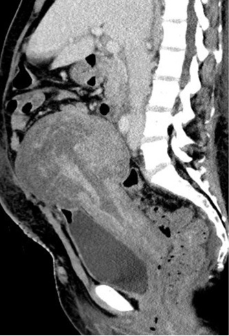
CT Abdomen with contrast – Sagittal
DIAGNOSIS
Uterine necrosis complicating B-lynch suture
IMAGING FINDINGS
Plain and contrast enhanced CT scan of abdomen shows an enlarged uterus with two paramedian vertically placed compression sutures. Non-enhancing myometrium at anterior and posterior uterine walls in between the sutures suggestive of ischemia. Plain CT scan of abdomen shows a hyperdense focus within the ischemic anterior uterine body, suggestive of hemorrhagic component. Sagittal and coronal reformatted contrast enhanced CT scan of abdomen shows non enhancing myometrium at the anterior and posterior uterine walls in between the sutures suggestive of ischemia.
DISCUSSION
This case demonstrated a rare but life-threatening condition complicating a common surgical procedure for primary postpartum hemorrhage. In this case, hysterectomy was subsequently performed due to persistent sepsis. Pathology confirmed hemorrhagic necrosis of the uterus with acute inflammation.
Primary postpartum hemorrhage is life threatening. Uterine compression suture techniques such as the B-Lynch suture is an effective surgical technique in the management of severe postpartum hemorrhage, and it is widely adopted due to simplicity of application and capacity of uterus preservation (1). Uterine necrosis, pyometra and uterine synechiae are the reported complications of this procedure (2). Uterine necrosis following B-Lynch is a rare but life-threatening condition (3-4), cross section imaging plays a vital role in diagnosis and prompt treatment with antibiotics and hysterectomy is necessary to prevent septicemia. Knowledge of this condition is vital for radiologists in making a correct diagnosis.
Degeneration of fibroids occurs can cause severe pain as well as fever or leukocytosis. Degeneration of fibroids results in a variable and often heterogeneous imaging appearance. Degeneration is categorized as hyaline, cystic, red, or calcific. Red degeneration is a hemorrhagic infarction of the fibroids, which is a well-known complication, especially during pregnancy secondary to insufficient blood supply and rapid growth associated with pregnancy. Red degeneration occurs in 8% of tumors complicating pregnancy (5).