Answer of February 2021
For completion of the online quiz, please visit the HKAM iCMECPD website: http://www.icmecpd.hk/
Clinical History:
This 57-year-old gentleman presented with acute onset of chest pain. The serum troponin was elevated to 400 ng/L (Reference range: < 40 ng/L)
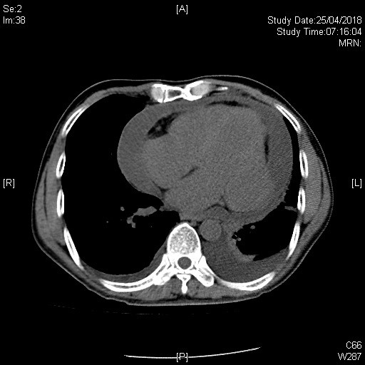
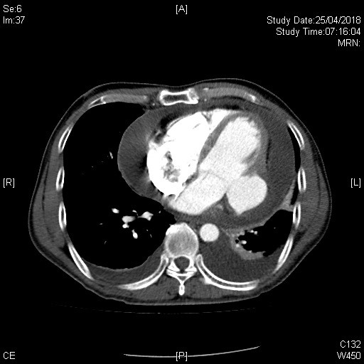
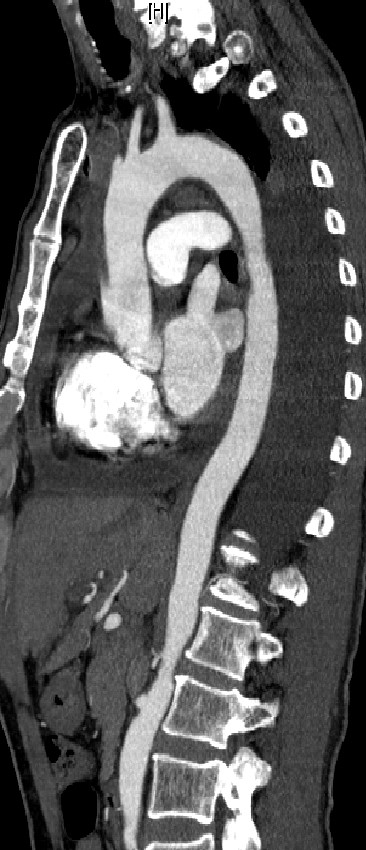
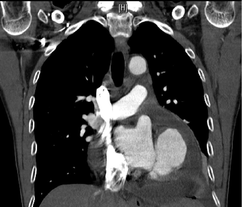
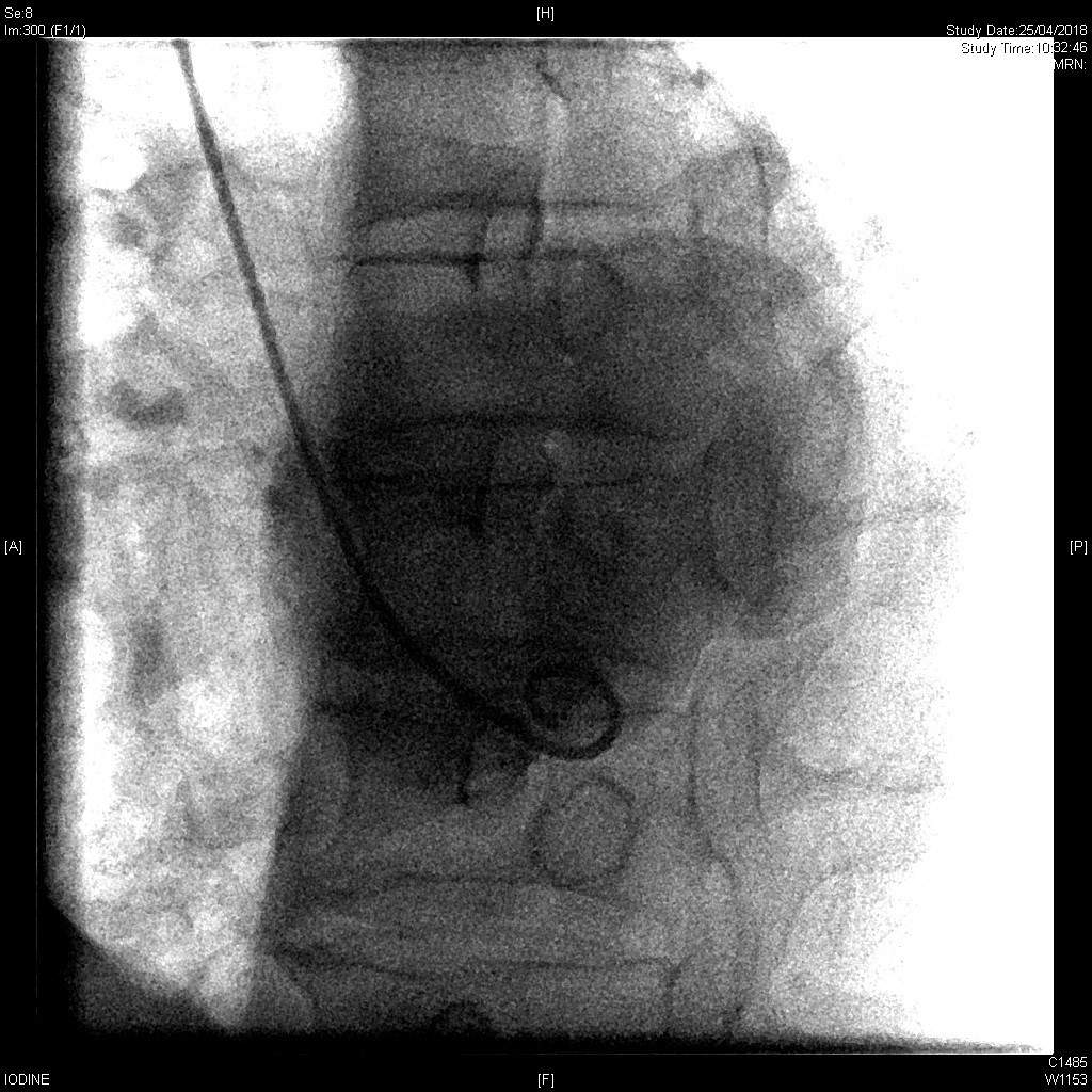
IMAGE FINDINGS:
CT and CTA
A contrast-enhanced wide-neck outpouching with lobulated contour at the lateral wall of the left ventricle [LV] of the heart connects with the cardiac chamber and measures around 4 cm in maximal dimension.
The neck of this outpouching was 2.5 cm.
Moderate hemopericardium (around 2.5 cm in thickness)
No intimal flap or aneurysm along the aorta.
Left ventriculogram: An outpouching at the LV lateral wall. No contrast extravasation.
DIAGNOSIS:
Left ventricular aneurysm.
DISCUSSION:
A left ventricular aneurysm is a discrete and dyskinetic area of the left ventricular wall with a broad neck. It is also known as a true aneurysm and develops in less than 5% of all patients with ST-elevation myocardial infarctions. [1] The clinical presentation may include heart failure but is non specific.
Features suggestive of a true ventricular aneurysm over a pseudoaneurysm is a wide neck The ratio of breach in the wall to the maximal diameter of the true aneurysm > 50% [2] is a suggestive finding. Cardiac MRI can be helpful for further evaluation of the cardiac walls and function in hemodynamically stable patients.