Answer of October 2020
For completion of the online quiz, please visit the HKAM iCMECPD website: http://www.icmecpd.hk/
Clinical History:
A 38 year-old lady underwent follow up MRI study of the lower abdomen after wide excision of an abdominal desmoid fibromatosis. An incidental finding was observed.
Imaging Findings
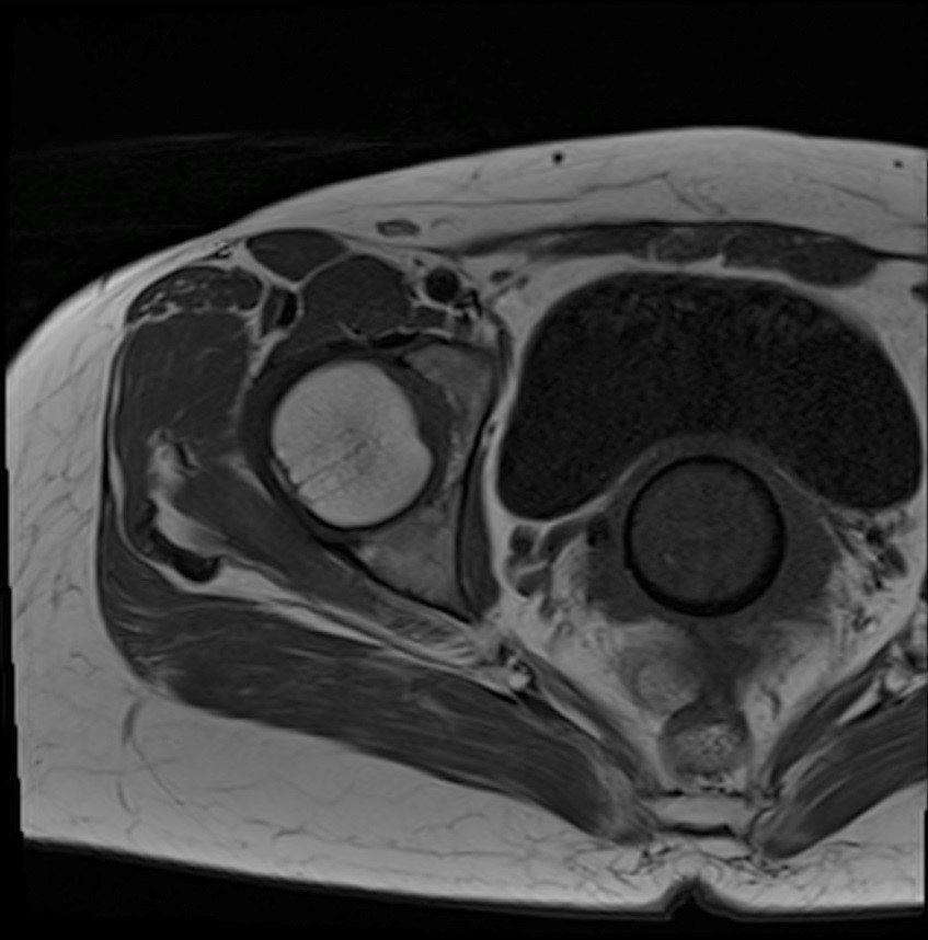
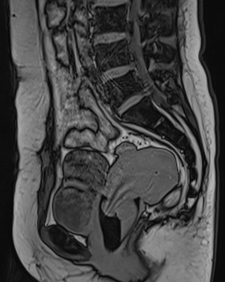
(Axial T1W, Sagittal T1W and T2W of the lower abdomen)
- No lesion in the right lower abdominal subcutaneous layer to suggest recurrence or residual tumour.
- Incidentally there is a flask-shaped structure in the vaginal vault, corresponding to a menstrual cup.
- T1-weighted isointense and T2-weighted hyperintense fluid within the cup, with layering of T1-weighted isointense and T2-weighted hypointense sediment at the dependent portion of the fluid, compatible with different ages of menstrual blood.
Discussion
Menstrual cup is an alternative sanitary product that can be used instead of tampons and sanitary pads during menstruation. It is increasingly prevalent especially in Western countries for its environmental-friendly advantages. It is a flask-shaped container which is placed into the vagina in order to collect the menstrual fluid. There are variations of capacities, ranging from 15mL to 45mL. Various brands are available but they all have the same shape. Although it is generally very safe to use with no increased risk of infection, there are case reports which describe urinary tract complications such as hydronephrosis in improperly positioned menstrual cups 1, as well as allergic reactions to the materials of the cup. Moreover, blood products within the menstrual cup could be mistaken for other significant pathologies in the pelvis such as a haematoma if the radiologist is not aware of this device.
Menstrual cups are made of MRI-compatible medical-grade silicon, natural gum rubber and thermoplastic elastomers, making most, if not all, of the cups safe to use during an MRI scan. It may partly obscure the vaginal canal or the uterine cervix. Thus, if urogenital system is the region of interest, the cup should be removed prior to the scan.
In conclusion, it is important for radiologists to be aware of this device and not to report it as pathology which could potentially cause unnecessary alarm to the patient and the physician.