Answer of March 2020
For completion of the online quiz, please visit the HKAM iCMECPD website: http://www.icmecpd.hk/
Clinical History:
This 23-year-old lady presented with right lower abdominal pain for a few months. Physical examination revealed a right lower quadrant mass and mild local tenderness.
 |
|
Image 1 |
 |
|
Image 2 |
 |
|
Image 3 |
 |
|
Image 4 |
 |
|
Image 5 (MIP) |
 |
|
Image 6 (Oblique image showing left hemi-pelvis) |
 |
|
Image 7 (Oblique image showing right hemi-pelvis) |
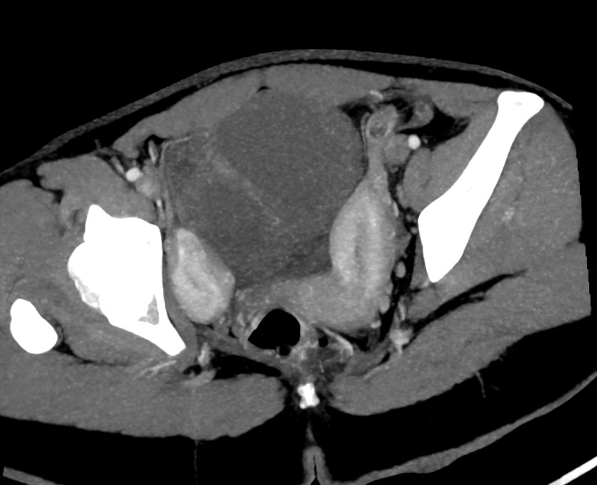
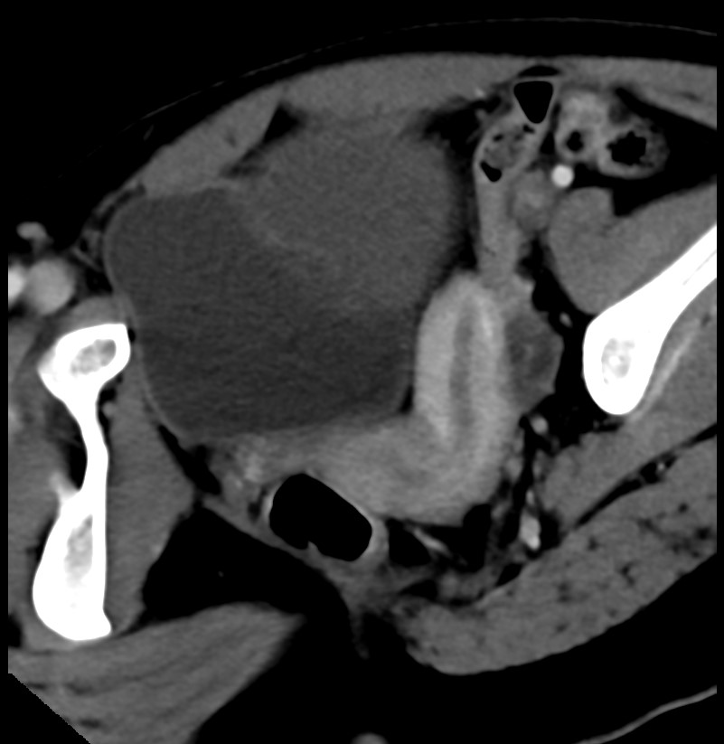
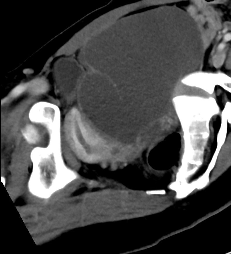
Imaging Findings:
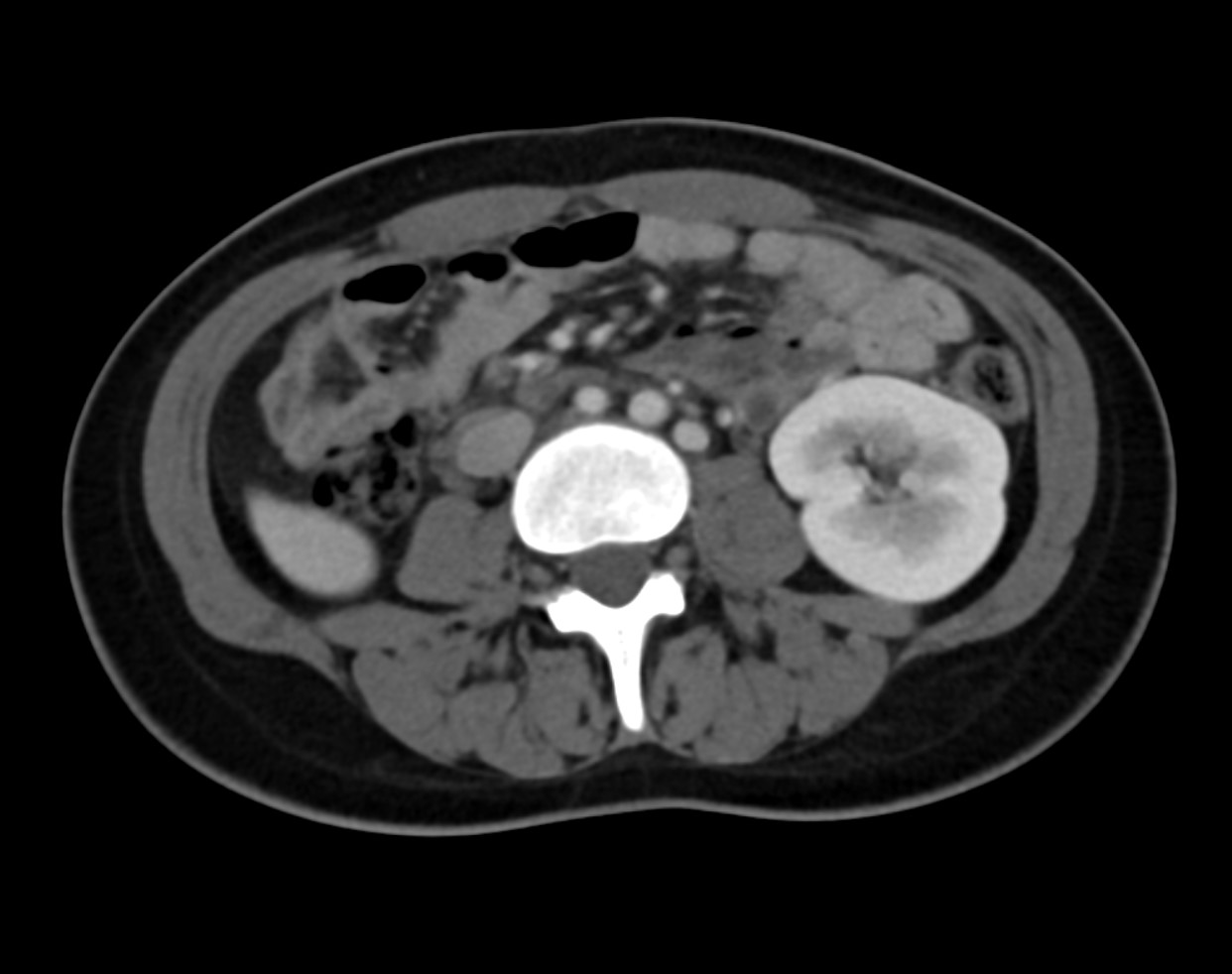
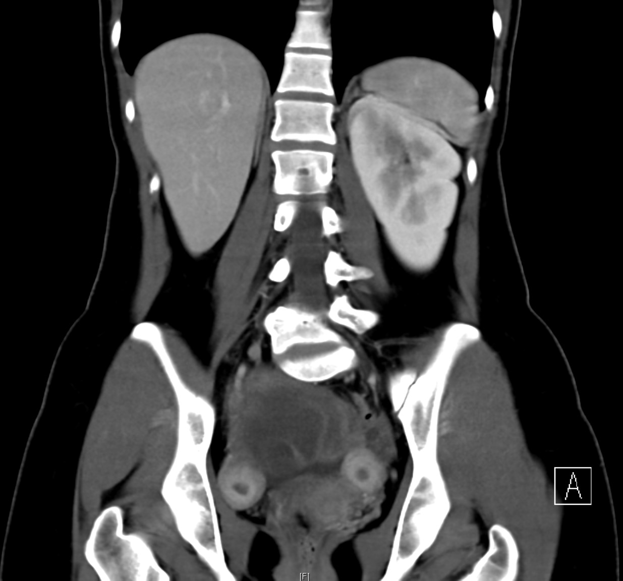
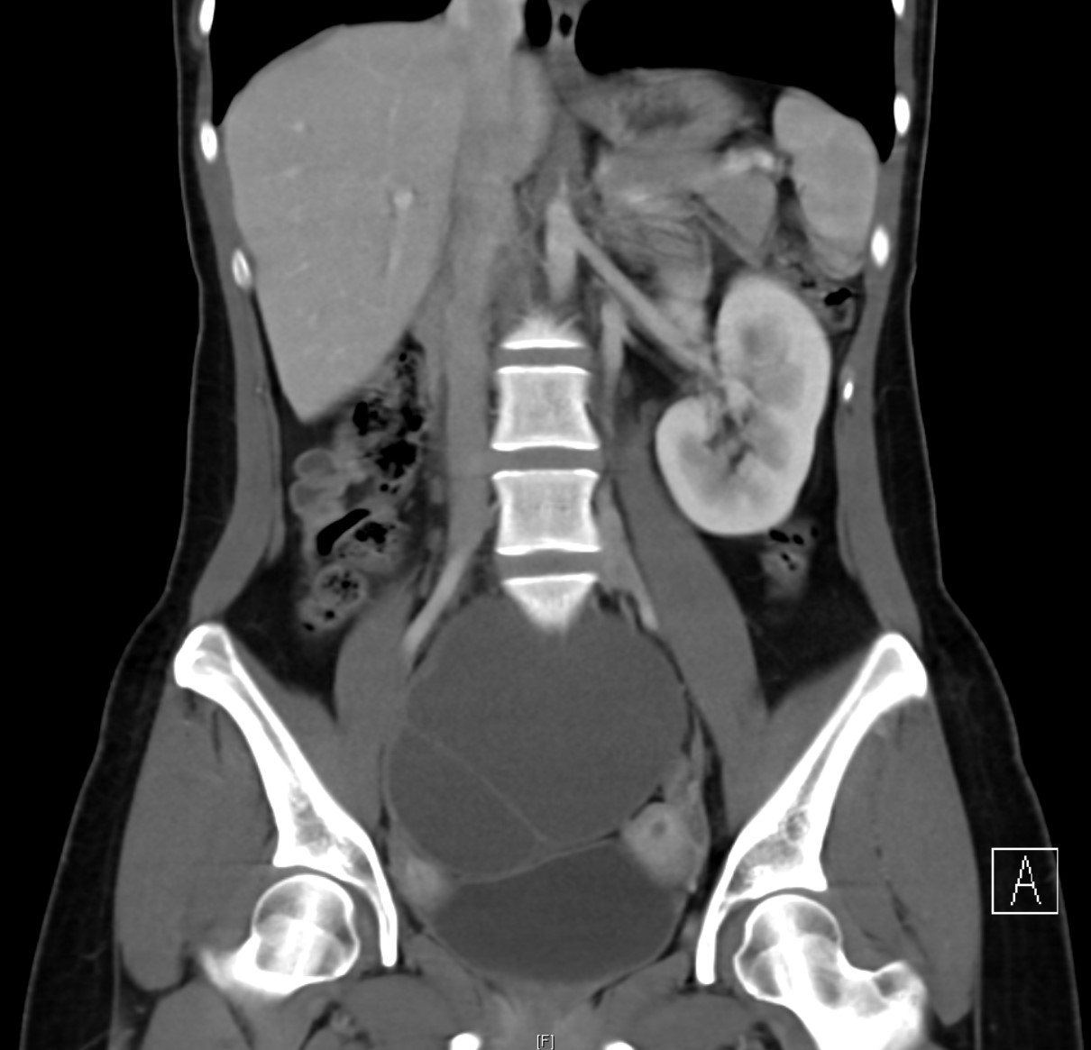
Normal right kidney is not seen in its expected location.
A large ~11cm structure with mixed cystic and soft tissue density in the right pelvic region represents right pelvic kidney with gross hydronephrosis. Paper-thin renal parenchyma indicates chronicity of the condition. The exact site of right collecting system obstruction is difficult to locate in the context of renal ectopia and unavailability of excretion phase images. The level of obstruction can be at the pelviureteric junction as a dilated right ureter is not seen.
Left kidney is unremarkable.
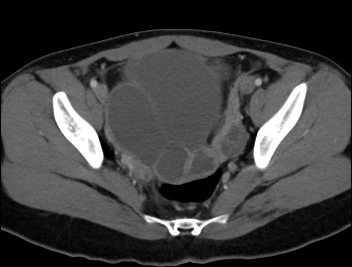
Abnormal configuration of the uterus is noted with a right rudimentary horn which contains enhancing endometrium. The left horn drains into a banana-shaped uterus curving to the left side. No connection between the right rudimentary horn and the uterine cavity is evident, suggestive of a non-communicating horn.
Diagnosis:
Left unicornuate uterus with right non-communicating rudimentary horn, associated with right pelvic kidney and chronic right hydronephrosis.
Discussion:
Unicornuate
uterus is a type of Müllerian duct anomaly (MDA), characterized by a
banana-shaped uterus usually draining a single Fallopian tube.
Its pathogenesis is due to a failure of one Müllerian duct to elongate while the other develops normally. While most patients are asymptomatic, some may suffer from cyclical lower abdominal pain due to obstructed rudimentary horn, recurrent miscarriage and infertility.
It can be classified into the subtypes according to presence/absence of contralateral rudimentary horn, the presence/ absence of rudimentary horn endometrium and whether the rudimentary horn is communicating with the main uterine cavity.
Unicornuate uterus is more commonly associated with renal anomalies than other types of MDA. Up to 40% of patients with unicornuate uterus are also found to have renal anomalies ipsilateral to the rudimentary horn, e.g. renal agenesis, pelvic kidney, cross fused renal ectopia, duplex kidney, etc.
Ultrasonography can be used as an initial imaging investigation to delineate the structural abnormalities of the genital tract, but it can be difficult to determine the type of MDA.
MRI is the modality of choice to assess the uterine configuration, myometrial zonal anatomy and presence of endometrial lining in the rudimentary horn. Imaging findings include curved and elongated uterus, banana-shaped external uterine contour, reduced uterine volume, asymmetric uterine configuration and a normal myometrial zonal anatomy.
Hysterosalpingogram can sometimes demonstrate the structural abnormality during workup for subfertility but the rudimentary horn cannot be fully assessed if it is non-communicating.
Co-existing renal anomalies should be actively screened for due to the high rate of association.
Surgical treatment is required for symptomatic patients suffering from severe cyclical abdominal pain, cryptomenorrhea, recurrent miscarriage, infertility and ectopic pregnancies.