Answer of December 2019
For completion of the online quiz, please visit the HKAM iCMECPD website: http://www.icmecpd.hk/
Clinical History:
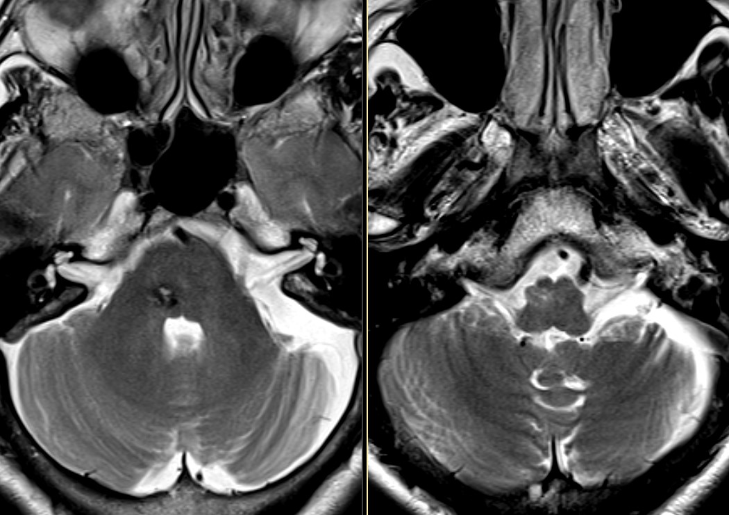
58 year old lady, history of brainstem haemorrhage with clot evacuation several years ago. Serial MRI was performed, with static appearance for the past number of years.
Discussion:
Hypertrophic olivary degeneration (HOD) is a unique type of transsynaptic degeneration resulting in hypertrophy of degenerated inferior olivary neurons rather than atrophy.
It results from damage to neuronal pathways in the Guillain-Mollaret triangle between the dentate nucleus of the cerebellum, red nucleus, and inferior olivary nucleus.
Palatal myoclonus is the classic clinical manifestation due to disruption of the central tegmental tract between the red nucleus and inferior olivary nucleus.
Most commonly occurs following focal lesions to the brainstem, such as hemorrhage, which in this case was related to a cavernous hemangioma.
Typically appears 4-6 months after brainstem insult and resolves by 3-4 years, though the T2 hyperintensity may persist longer.