Answer of February 2019
For completion of the online quiz, please visit the HKAM iCMECPD website: http://www.icmecpd.hk/
Clinical History:
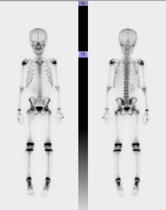
This is a 9-year-old boy with recent diagnosis of Clear cell sarcoma of left kidney referred for bone scintigraphy. Patient has no symptom of bone pain.
 |
|
Image 1 of 2 |
 |
|
Image 2 of 2 |
Imaging Findings:
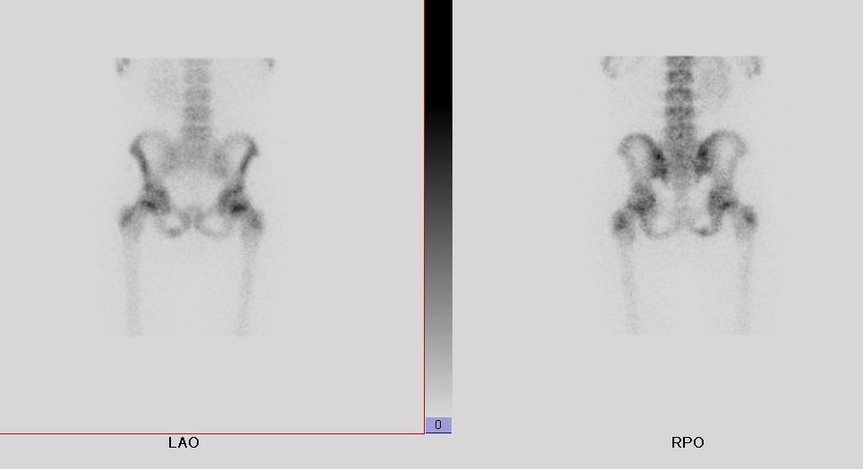
- Increased bone tracer uptake at the right ischiopubic junction
- The contralateral side shows no increased uptake
- The rest of the skeletal uptake appears normal for age
- Absent of left renal tracer activities is consistent with left nephrectomy
- Urine contamination is noted at the right groin region but is cleared after cleaning the groin region before the regional views were obtained
Diagnosis:
right ischiopubic synchondrosis, a normal finding.
Discussion:
Ischiopubic synchondrosis is the cartilaginous junction between distal end of the inferior pubic ramus and the distal end of the inferior ischial ramus and acts similar to a primary ossification center. It ossifies between ages of 4 to 12 years. Asymmetry is common and presents as uneven swelling, in particularly around age of 8 years. The presence of immature osteoid and developing hydroxyapatite crystals explains for the uptake of bone tracer such as Tc-99m methylene diphosphonate (MDP). Before fusion there may be some mild demineralization and swelling at the ischiopubic synchondrosis. The intensity of uptake at the ischiopubic synchondrosis can be similar to that at the area of growth plate.
In this case, the focal uptake in the typical location is a normal physiological finding and correlated radiographically with incomplete fusion of right ischiopubic synchondrosis. The importance of recognising this is to avoid unnecessary imaging or intervention, as it is a common finding in paediatric bone scintigraphy. However, it is worthwhile to note that focal uptake in a bone scintigraphy can otherwise be a non-specific finding with many possible causes, such as osteomyelitis, healing fracture and metastasis, for example, in particular if patient is symptomatic.
It has been reported that ischiopubic synchondrosis is FDG-avid on F-18 FDG PET/CT before fusion of the ischial and pubic bone.
Radiographic abnormalities of ischiopubic synchondrosis include lucencies, bony enlargement, and irregular mineralization have been incidentally noted during this fusion process.