Answer of January 2019
For completion of the online quiz, please visit the HKAM iCMECPD website: http://www.icmecpd.hk/
Clinical History:
A 52 year old lady complained of abdominal pain, vomiting and reduced bowel opening. History and physical examination findings were non-revealing. An abdominal radiograph, CT scan of the abdomen and colonoscopy were arranged.
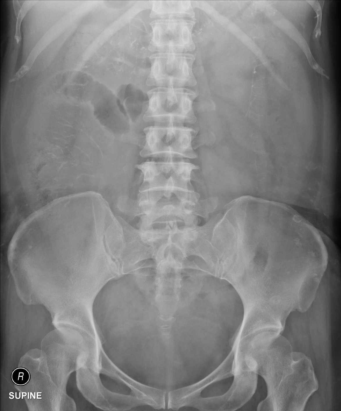
Fig. 1. Supine abdominal radiograph
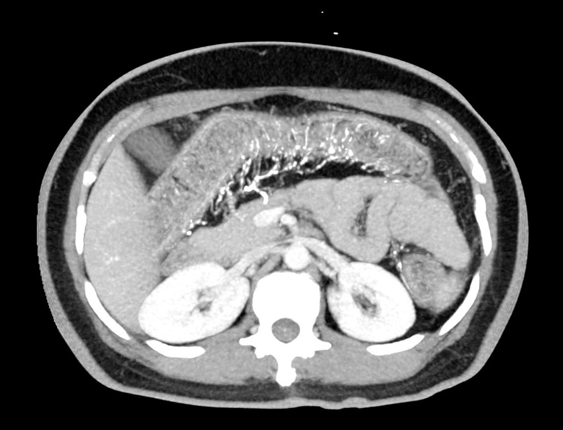
Fig. 2. Selected image from axial reformatted CT with MIP
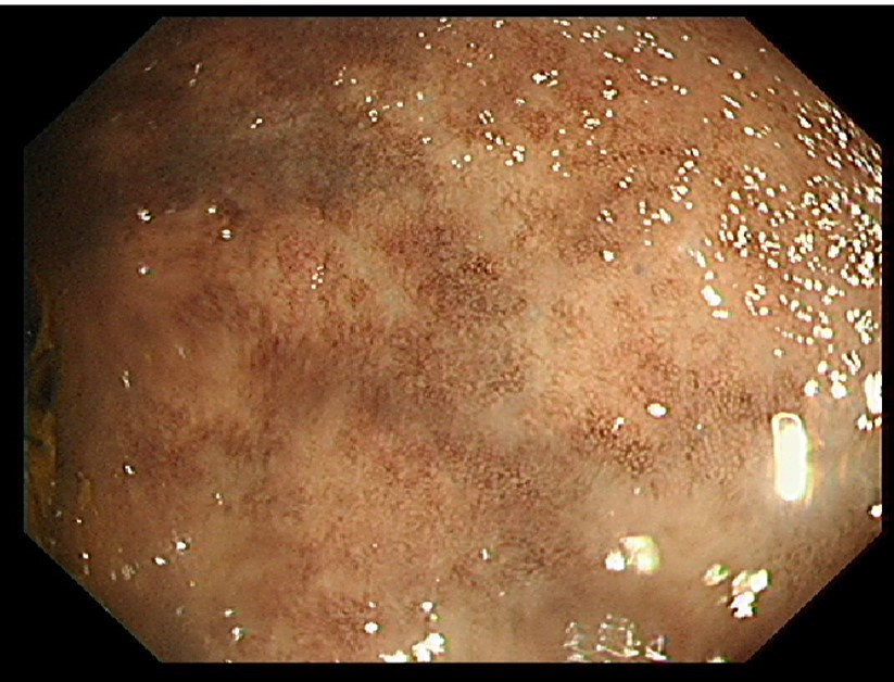
Fig. 3. Endoscopic image of the transverse colon
Imaging Findings:
- Extensive thread like calcifications extending radially along the large bowel mesentery, with predominant R sided involvement.
- Thumbprinting of the transverse colon.
- Prominent small bowel loops.
- Brownish discoloration and loss of vascular pattern of the transverse colon mucosa, suggesting chronic ischaemic colitis.
Discussion:
Ischaemic bowel disease is more commonly caused by arterial atherosclerotic disease than venous insufficiency, in which case thrombotic occlusion at a proximal level eg portal vein or SMV is the common aetiology.
Mesenteric phlebosclerosing colitis is a rare entity characterized by diffuse calcifications and occlusion of the large bowel distal venous supply resulting in ischaemic colitis.
It was coined in 1997 by the Japanese pathologist Iwashita and colleagues, who presented seven cases of chronic ischaemic colitis with extensive calcifications of the mesenteric veins and their tributaries without associated thrombotic or inflammatory phenomenon. Since then, <100 cases have been reported, with most patients being Japanese (75%) or Asians. Ingestion of toxic herbal medicine was posited as a cause, though larger scale studies are required to verify this hypothesis.
The presenting age ranges from 33-77 years old, with males and females equally affected. Symptoms are typically chronic and non specific such as abdominal pain, vomiting and diarrhoea. There can be acute exacerbation caused by functional ileus. Often the diagnosis is first suspected radiologically and subsequently confirmed by histology. More than half of the patients require colectomy which is considered a curative treatment.