Answer of May 2017
For completion of the online quiz, please visit the HKAM iCMECPD website: http://www.icmecpd.hk/
Clinical History:
This 53 year old gentleman with no relevant past medical history was referred for persistent painful swelling of the left ring finger for 4 months. He reports an episode of contusion 3 years ago.
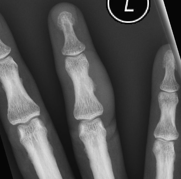
Figure 1.
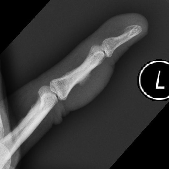
Figure 2.
Diagnosis:
Bizarre parosteal osteochondromatous proliferation (Nora lesion)
Imaging Findings:
Radiographs showed a sclerotic, cortically based lesion at the radial-dorsal aspect of the diaphysis of the intermediate phalange, with ill-defined margins. No involvement or continuity of the medulla is noted. Overlying subcutaneous soft-tissue thickening is present.
Discussion:
Bizarre parosteal osteochondromatous proliferation (BPOP, a.k.a Nora lesion) is a rare benign condition, of locally aggressive and often recurrent osteochondromatous exostosis originating in the cortex. It presents a diagnostic challenge to radiologists, physicians and surgeons alike. The aetiology, natural history and clinical course of BPOP is not completely understood.
Over 50% presents as a painful growth from the small bones of the hands and feet, although they can less commonly occur in the long bones and rarely also in the skull, maxilla, or sesamoid bones.
Lesions typically present as a slowly enlarging swelling in the 3rd to 5th decades with a mean of 34 years, with no gender predilection. Pain is present in about half of cases. About half of patients report prior trauma at the site of occurrence, although definitive inciting factors are yet to be determined.
Classic imaging appearance is an exophytic bony ossification or calcification, contiguous with (without disrupting) the bony cortex on radiographs. Unlike osteochondromas, there should be no extension or continuity with the bony medullary cavity.
Main differential considerations includes parosteal osteosarcoma, the most important condition to exclude, although this generally occurs in the long bones especially the distal femur, and only rarely involves the small bones of the hands and feet where BPOP tends to occur. Myositis ossificans is another differential, but generally occurs in association with larger muscles and has a centripetal pattern of ossification. Periosteal chondromas exhibits a characteristic saucerisation (scalloping) of the underlying cortex. Turret exostosis and florid periostitis are related reactive processes of the bone that can appear similar to BPOP radiographically, and have been proposed to reflect a continuous spectrum of diseases along with BPOP. Biopsy typically gives mixed osseous, cartilaginous and fibrous tissue elements, sometimes with presence of a cartilage cap.
BPOP should be considered in cortically based lesions of the small hand bones as in the presented case. While radiographs alone are sufficient for diagnosis in presence of typical radiographic and clinical features, but its often aggressive and non-specific appearance can pose a diagnostic challenge to radiologists and clinicians. A multidisciplinary approach will likely be most beneficial to management and patient outcome.

