Answer of December 2016
For completion of the online quiz, please visit the HKAM iCMECPD website: http://www.icmecpd.hk/
Clinical History:
A 72-year-old lady complains of headache and blurring of vision for 3 months. There was no focal neurological sign elicited by clinician. MRI brain was performed to investing for the symptoms.
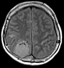
Proton-Density weighted axial
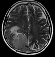
T2 weighted axial
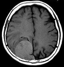
T1 weighted axial
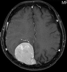
T1 weighted axial, post-gadolinium
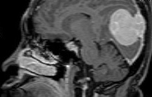
T1 weighted sagittal, post-gadolinium
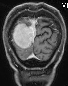
T1 weighted coronal, post-gadolinium
DWI axial
Diagnosis:
Meningeal haemangiopericytoma
Discussion:
Meningeal haemangiopericytomas are rare tumours of the pericytes that originate in the meninges, which contributes to 1% of all central nervous system tumours, and can be difficult to be differentiated from meningiomas, which are far more common. When compared with meningiomas, meningeal haemangiopericytomas have a more aggressive nature, high rate of local recurrence and propensity for late, extra-cranial metastases.
Both haemangiopericytomas and meningiomas are dural-based lesions and are often hyperdense on CTs. There are several features that may enable differentiation of haemangiopericytomas from meningiomas. Haemangiopericytomas are not associated with calcifications, unlike meningiomas. Haemangiopericytomas often appear more lobulated than meningiomas. Rather than hyperostosis, which is often seen with meningiomas, haemangiopericytomas show bone erosions in more than half of the cases and do not show hyperostosis. About one-third of haemangiopericytomas demonstrate a narrow base of dural attachment, while the remaining two-thirds show a broad-based attachment with a dural tail sign. Haemangiopericytomas typically show heterogeneous enhancement, in contrast to the homogeneous enhancement often seen with meningiomas. Haemangiopericytomas often show prominent internal serpentine vessel voids.
The differentiation of haemangiopericytomas from meningiomas is helpful in prediction of the prognosis and the management plan, which may require pre-operative embolization to reduce intra-operative blood loss, and adjuvant radiotherapy after total excision to reduce the much higher local recurrence rate.
Other less common dural lesions that may mimic meningiomas include solitary fibrous tumours, gliosarcoma, leiomyosarcoma, dural metastases, plasma, neurosarcoidosis.
