Answer of October 2016
For completion of the online quiz, please visit the HKAM iCMECPD website: http://www.icmecpd.hk/
Clinical History:
A 44 year old lady presented with vaginal bleeding.
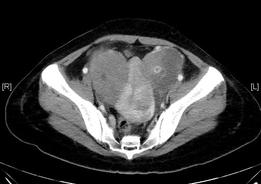
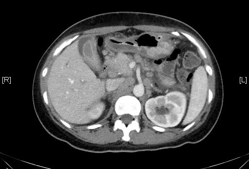
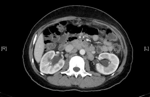
Axial CT scan
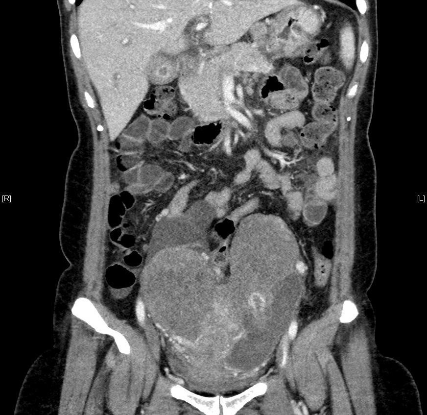
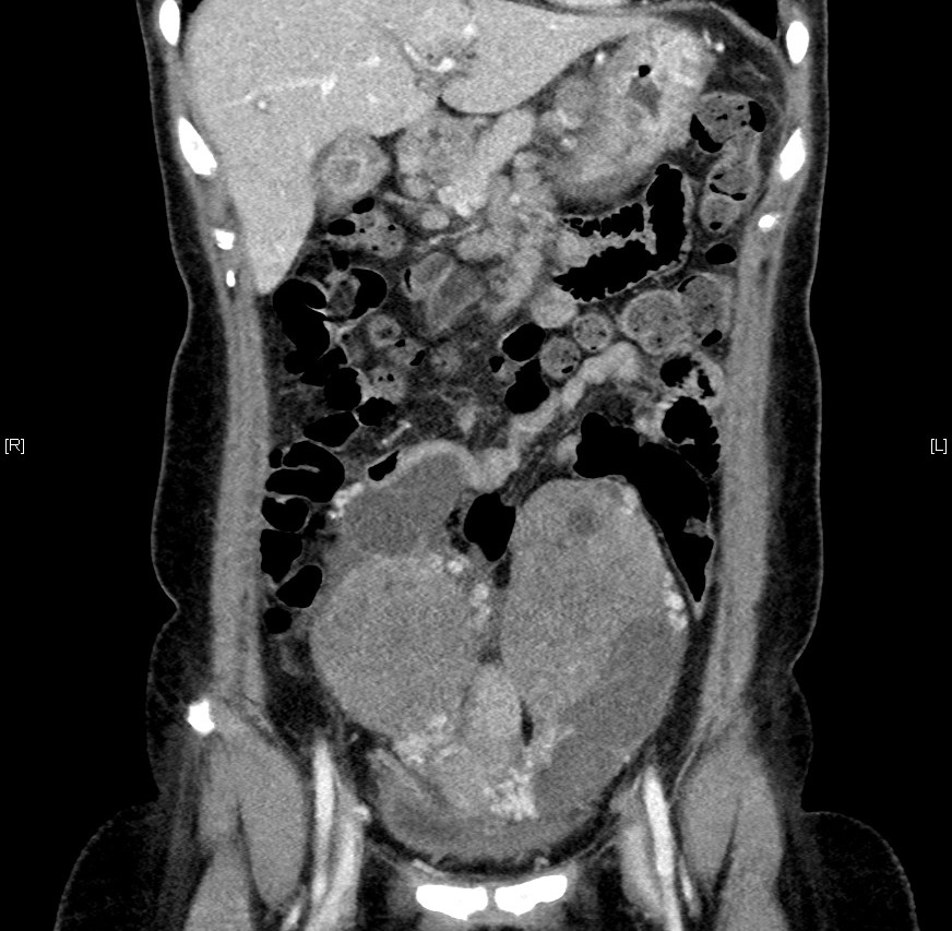
Coronal CT scan
Diagnosis:
Krukenberg tumor
Discussion:
Krukenberg tumor is a metastatic tumour to the ovaries, most of them contain mucin-secreting signet ring cells. Colon and stomach are the most common primary tumours to result in ovarian metastases, followed by the breast, lung, and contralateral ovary. In 20% of case, Krukenberg tumor antedates the discovery of the primary tumor. It is most common in age 50-60 female. Imaging findings mainly reveal bilateral ovarian complex mass with solid component with cystic degeneration.
MRI pelvis could be helpful in making diagnosis. It will demonstrate hypointense solid components (dense stromal reaction) and internal hyperintensity (due to presence of mucin) on T1- and T2-weighted MR images.
Differentiation between primary and metastatic ovarian carcinoma is of great importance with respect to treatment and prognosis. Metastatic ovarian carcinoma is commonly associated with ascites. Histological diagnosis may be necessary in equivocal case.
In this case, there is marked lymphadenopathy in the peri-gastric region and upper retroperitoneal region. There is also mild thickening along the gastric antrum. This is highly suspicious with gastric cancer which was subsequently proven as poorly differentiated adenocarcinoma of stomach with endoscopic biopsy. Bilateral hydronephrosis was also found due to the obstruction from pelvic masses.
