Answer of February 2016
For completion of the online quiz, please visit the HKAM iCMECPD website: http://www.icmecpd.hk/
Clinical History:
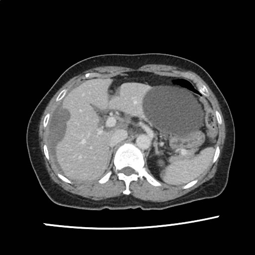
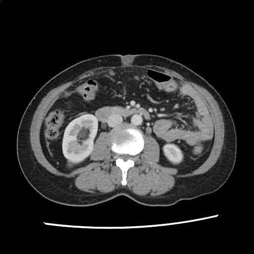
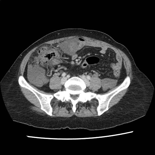
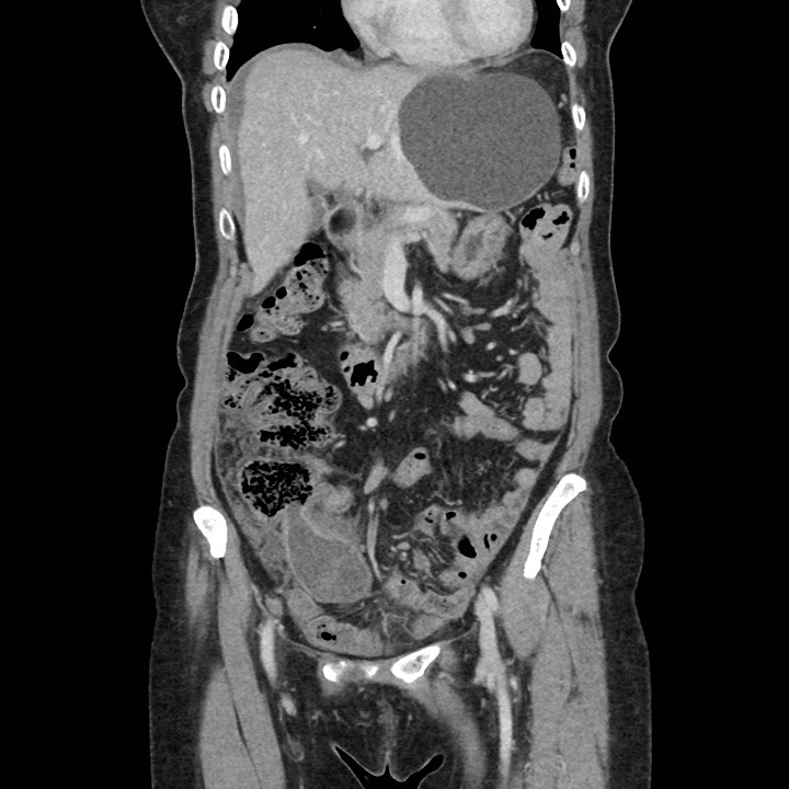
A 56 year-old female patient presented with blood stained sputum. A CT thorax was initially performed and the lung was found to be normal. However, incidental finding in the upper abdomen was detected and a contrast CT scan of abdomen and pelvis was subsequently carried out.
Figure 1
Figure 2
Figure 3
Figure 4
Diagnosis:
Pseudomyxoma
peritonei due to mucinous adenocarcinoma of appendix
Discussion:
CT scan shown loculated hypodense collections with the abdominal and pelvic cavities with scalloping of hepatic and splenic surfaces. The appearance is compatible with pseudomyxoma peritonei. The collections have no rim enhancement to suggest abscess formation. The appendix is grossly distended with hypodense material with normal wall thickness, suggestive of a mucocele. A Sister Mary Joseph nodule is seen representing umbilical metastasis and signifying underlying malignant process. The most likely unifying diagnosis is pseudomyxoma peritonei secondary to mucinous adenocarcinoma of the appendix.
Patient underwent surgery and the diagnosis of mucinous adenocarcinoma of the appendix was confirmed histologically. The umbilical nodule was also shown to be involved.
Pseudomyxoma peritonei refers to accumulation of a large amount of mucinous or gelatinous material over peritoneal surfaces. Patients usually present with increasing abdominal girth and distension, abdominal pain and weight loss. It is commonly accepted that majority of the cases are caused by low-grade mucinous carcinomas arising in the appendix that ruptures or penetrates into he peritoneal cavity. Less commonly, the condition can be caused by peritoneal mucinous carcinomatosis of the gastrointestinal tract, gallbladder, pancreas or ovary. Pseudomyxoma peritonei is a rare condition with an incidence of 1 in 100000 per year. It is more commonly seen in middle age female. Abdominal radiograph has no role in diagnosis and CT is usually the imaging modality of choice.
Scalloping of visceral surfaces of the liver or spleen is an important diagnostic feature that helps to differentiate the condition from simple ascites. It is due to extrinsic mass effect of the mucinous collection.
Complication of bowel obstruction may be seen in some advanced cases.
Treatment usually consists of surgical debulking. Intraperitoneal chemotherapy may be considered in individual cases.


