Answer of November 2015
For completion of the online quiz, please visit the HKAM iCMECPD website: http://www.icmecpd.hk/
Clinical History:
A 14 year old lady presented with on and off fever associated with cough and sputum for 1 month. She then experienced increased in shortness of breath and chest pain and attended the accident and emergency department. There was no haemoptysis and no weight loss. White cell count was elevated and rest of the blood results were unremarkable. A chest radiograph and CT thorax was performed.
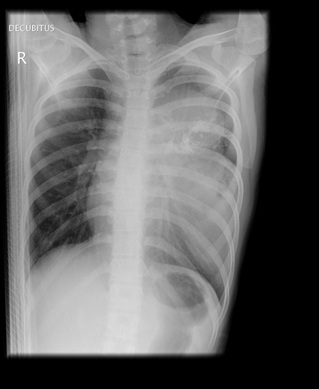
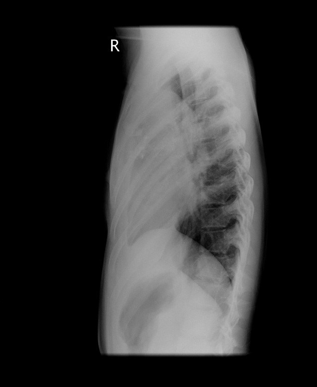
CXR
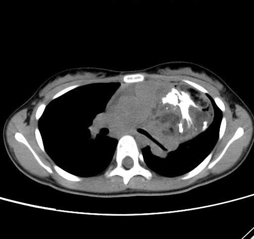
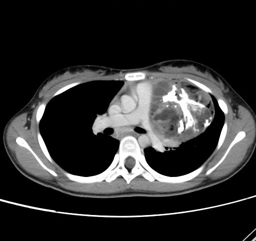
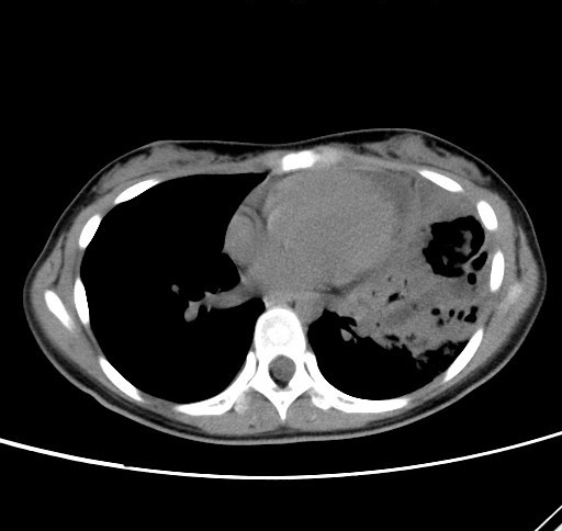
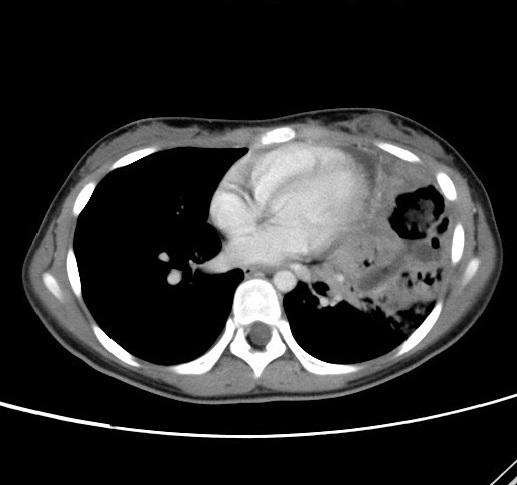
CT Thorax Non Contrast Post Contrast
Diagnosis:
Mediastinal teratoma complicated by rupture
Findings:
CXR: Large lobulated mass occupying left hemithorax with internal calcification, confirmed to be located within the anterior mediastinum on lateral view.
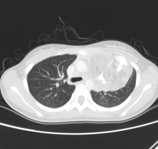
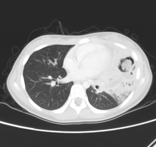
CT Thorax: Anterior mediastinal mass with internal fat density and calcification, suggestive of teratoma. Irregularity is noted at inferior border of the tumor with air locules seen at the inferior aspect of the lesion. It is associated with consolidation at the adjacent lingular segment of left lung and fat density within the consolidation suggestive of rupture and lipoid pneumonia.
Discussion:
Differentials of anterior mediastinal masses included thyroid lesions, thymic masses, teratoma and lymphoma. The most common primary germ cell tumors in the mediastinum are mature cystic teratomas containing ectodermal, mesodermal, and endodermal derivatives. These patients are usually asymptomatic until the mass is sizeable and compresses the mediastinal structures or rarely, rupture. Some authors have proposed that large size, infection and sebaceous materials/digestive enzymes leading to inflammation increase the chance of rupture. When rupture occurs, patients may complain of increase in dyspnea, chest pain and hemoptysis. If low attenuation fat density is present within the adjacent consolidation then rupture with lipoid pneumonia needs to be considered. Complete surgical resection is the mainstay of treatment, but in immature cases with malignant potential then chemotherapy or radiation therapy may be indicated.
