Answer of July 2015
For completion of the online quiz, please visit the HKAM iCMECPD website: http://www.icmecpd.hk/
Clinical History:
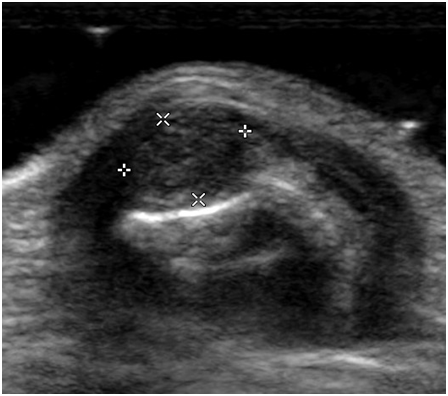
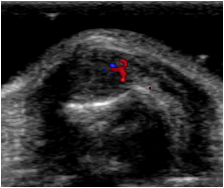
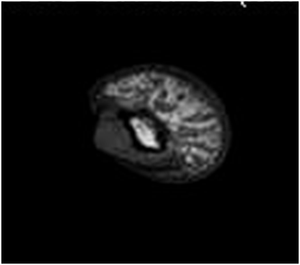
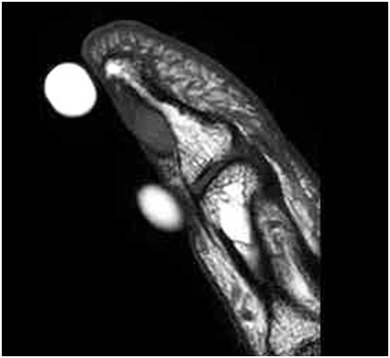
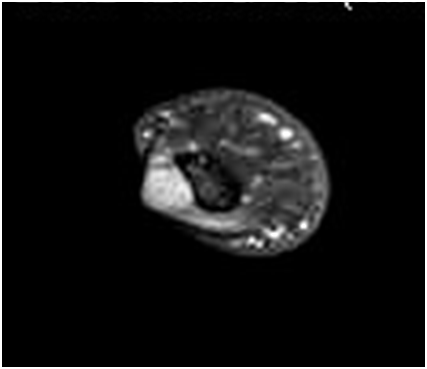
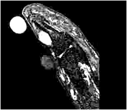
A 36 year-old woman presented with left thumb pain over the nail bed for over 10 years with hypersensitive to weather change or upon touching. Physical examination showed a raised red swelling over left thumb nail with tenderness on palpation. Ultrasound (USG) of the nail bed region was shown (2 images). Magnetic resonance imaging (MRI) was also performed (4 images – upper row: axial T1 weighted image, sagittal T1 weighted image; low row: post-contrast fat-suppressed axial T1 weighted image, fat-suppressed sagittal T2-weighted image)
USG
MRI
DIAGNOSIS:
Glomus tumor
DISCUSSION:
Glomus tumor is a rare benign vascular neoplasm that arises from the glomus body, which is an arteriovenous shunt within the dermis that contributes to temperature regulations. It accounts for 1% to 4.5% of tumors in the hand. The glomus body is more concentrated in the digits, particularly in the subungal germinal matrix. It is more common in females in the 30-50 year-old. They can be multiple in ~10% of the cases. Classic clinical triad of fingertip pain, tenderness, and sensitivity to cold has been described. Delays in diagnosis are not uncommon with symptoms persist for several years before an actual diagnosis is made. It can present as a small red-blue nodule under the finger nail due to previous hemorrhage.
USG typically shows a subungual hypoehocic lesion, which demonstrates hyper-vascularity on doppler interrogation. MRI is invaluable to make the diagnosis and provide exact location of the lesion. Glomus tumor typically has low to intermediate signal on T1-weighted images, high signal in T2-weighted images, and shows avid contrast enhancement due to vascularity. On T1-weighted images, hyperintensity in some of the tumors is due to hemorrhage or due to large vascular channels. Contrast-enhnaced MR angiography may be used to demonstrate the characteristic vascular nidus .
The differential diagnosis may include mucoid cyst, haemangioma and epidermoid inclusion cyst.
Mucoid cyst is simply a ganglion that classically located in the dorsal aspect of the distal interphalangeal joint. This lesion follows the signal intensity of fluid on all pulse sequences and do not enhance following contrast administration.
Haemangioma has similar imaging features to those of a glomus tumor. It is however more superficial in location in the papillary dermis and the epidermis.
Epidermoid inclusion cyst can appear similar to glomus tumor in imaging characteristics. It is well defined and of T2 hyperintensity. The key to the diagnosis is the clinical history and presentation. It results from prior penetrating trauma with subsequent proliferation of the implanted dermal elements, and it is usually painless.
Treatment for glomus tumor is surgical resection, with low likelihood of recurrence.
