Answer of January 1999
Clinical History:
F/63. Incidental finding on CXR. CT performed.
Figure 1 CXR (PA)
Figure 2 CXR (lat.)
Figure 3 Plain CT thorax
What is your diagnosis?
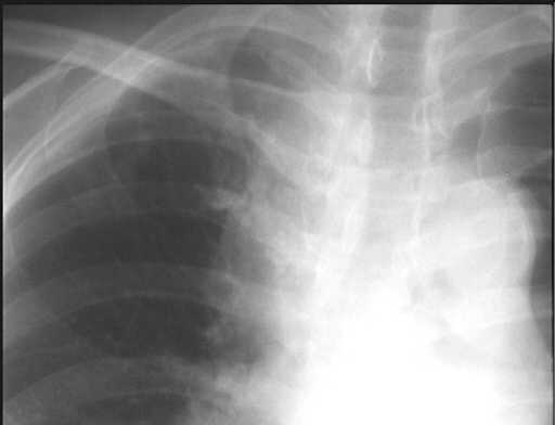
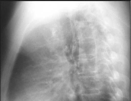
Figure 1 Figure 2
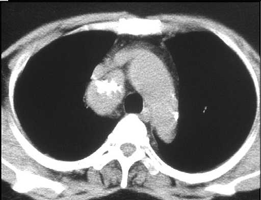
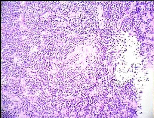
Figure 3 Figure 4
Diagnosis:
CASTLEMAN DISEASE
Discussion:
Figure 1 & 2 CXR (PA & lat.) showed R superior mediastinal mass with calcification.
Figure 3 CT showed calcified R superior mediastinal mass behind SVC.
Figure 4 Mediastinoscopy & biopsy with histology confirming Castleman disease, hyaline vascular type.