Answer of July 1999
Clinical History:
M/16 Right hip pain for few months.
Figure 1 Pelvic x-ray
Figure 2 Right hip x-ray
What is your diagnosis?
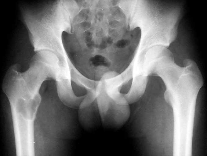
Figure 1
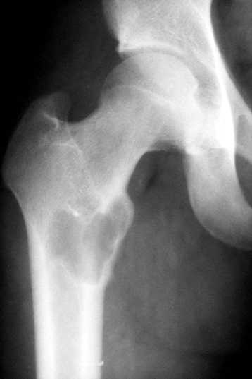
Figure 2
Diagnosis:
Giant cell tumour
Discussion:
Figure 1 Pelvic x-ray shows a lucent lesion in proximal right femur, extending to the tip of lesser trochanter with cortical thinning and mild bony expansion seen.
Figure 2 Right hip x-ray demontrates a narrow margin of transition with a punch out margin , minimal border sclerosis and slight periosteal reaction. Biopsy revealed Giant cell tumour.