Answer of October 1999
Clinical History:
M/67 History of diabetes mellitus. Admitted with RUQ pain and fever.
Figure 1 AXR
Figure 2 AXR(RUQ,36 hours later)
What is your diagnosis?
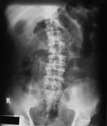
Figure 1
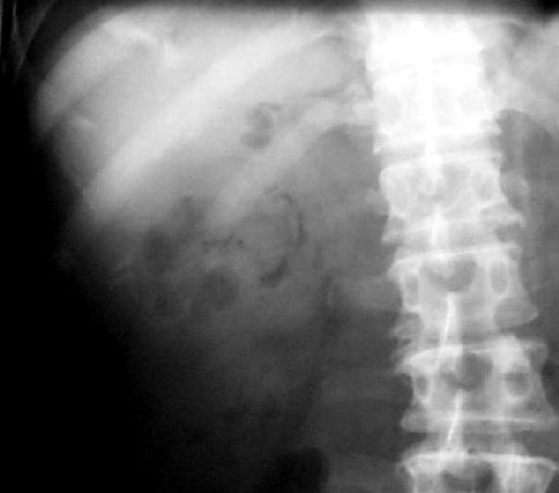
Figure 2
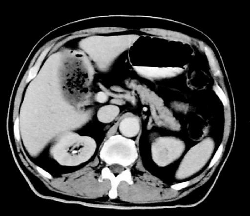
Figure 3
Diagnosis:
Emphysematous cholecystitis
Discussion:
Figure 1 AXR shows collection of gas with the configuration of gall bladder in the right upper quadrant.
Figure 2 AXR(RUQ, 36 hours later) demonstrates that there is now mottled and curvilinear gas lucencies in the gall bladder region.There is also gas in the cystic duct.
Figure 3 CECT abdomen shows that there is gas in the gall bladder lumen and in the thickened gall bladder wall chacteristic of emphysematous cholecystitis.