Answer of November 1999
Clinical History:
M/60 Bilateral lower limbs weakness
Figure 1 NECT brain
Figure 2 NECT brain
Figure 3 CECT brain
What is your diagnosis?
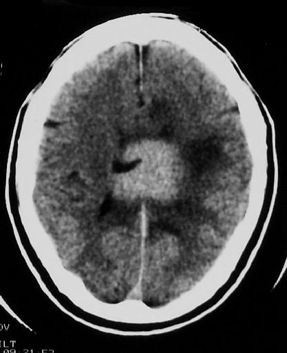
Figure 1
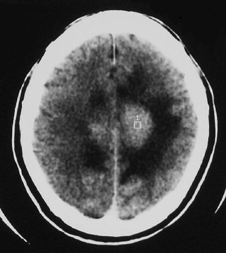
Figure 2

Figure 3
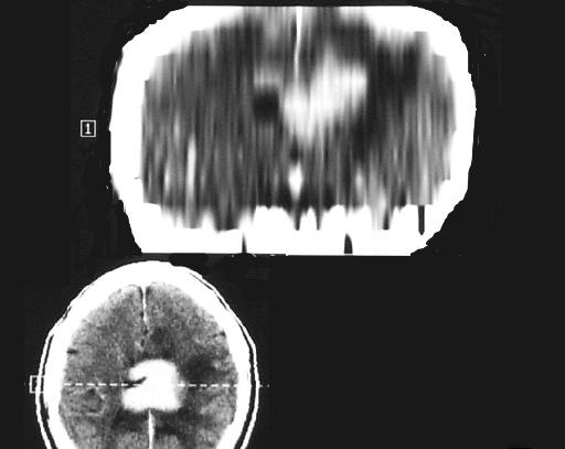
Figure 4
Diagnosis:
Primary cerebral lymphoma
Discussion:
Figure 1 NECT brain shows a hyperdense mass in the corpus callosum and adjacent peritumour oedema.
Figure 2 NECT brain shows the mass to be seperated from the falx.
Figure 3 CECT brain demontrates the strong and homogenous contrast enhancement of the mass.
Figure 4 Coronal reformat image of the brain clearly shows the mass to be in the corpus callosum. Biopsy revealed lymphoma of brain.