Answer of August 2001
Clinical History:
F/ 80. Sudden onset of aphasia
Figure 1-2 Axial T2-weighed scan of brain
Figure 3-4 Axial diffusion weighted image of brain
Figure 5-6 Axial ADC image of brain
What is your diagnosis?


Figure 1 Figure 2
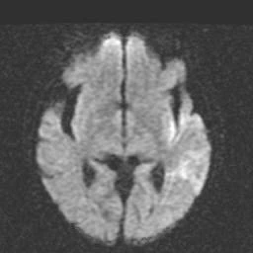
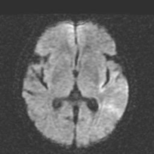
Figure 3 Figure 4
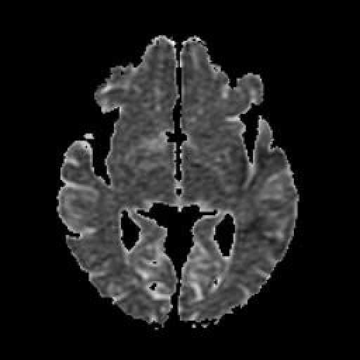

Figure 5 Figure 6
Diagnosis:
Hyperacute (less than 1 day) cerebral infarct.
Discussion:
Figure 1 & 2 Axial T2W scan of brain showing no abnormality.
Figure 3 & 4 Diffusion Weighed axail scan of brain showing abnormal signallesions at left temporo-occipital lobe.
Figure 5 & 6 ADC scan of brain showing low signal intensity in the corresponding sites.