Answer of October 2001
Clinical History:
F/ 74. c/o Shortness of Breath
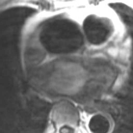
Figure 1 SE Axial T-1 weighed heart
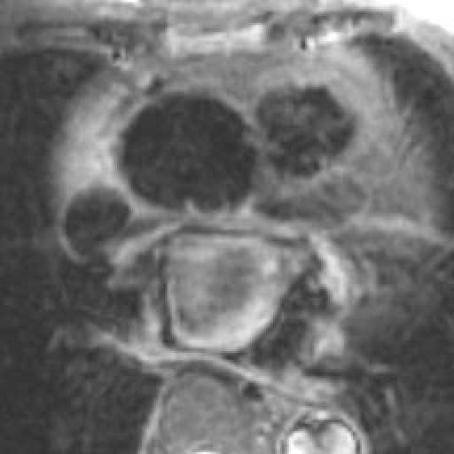
Figure 2 FSE Axial T-2 weighed heart
Figure 3 Parasagittal CINE heart
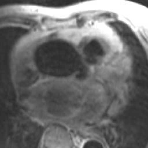
Figure 4 SE Post-contrast axial heart
Figure 5 CINE
What is your diagnosis?

Diagnosis:
Thrombophlebitis Migrans secondary to CA ovary.
Discussion:
Figure 1 & 2 Axial post-contrast scan of calf showing abnormal filling defects in the deep veins of both lower limbs.
Figure 3 Coronal post-contrast axial scan of calf showing thrombus in deep veins of both lower limbs.
Figure 4 axial scan of pelvis showing abnormal soft tissue mass behind uterus. During operation the mass is confirmed to be carcinoma of ovary.