Answer of March 2002
Clinical History:
Asthma with repeated hospital admission.
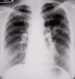
Figure 1 Chest Radiograph
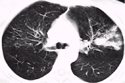
Figure 2 High resolution CT scan of lungs
Figure 1
Figure 2
Diagnosis:
Allergic bronchopulmonary aspergillosis.
Figure 1 Chest Radiograph
Fingers-in-glove shadows in the left upper and middle zones suggesting bronchocoeles. Reticular shadows in the right upper and middle zones.
Figure 2 High resolution CT scan of lungs
Left upper lobe bronchocoeles are seen. Right upper lobe fibrosis is seen.

