Answer of April 2002
Clinical History:
M/74 Upper back pain for a few days.
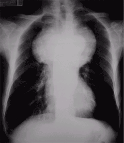
Figure 1 Chest Radiograph
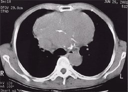
Figure 2 CT thorax (noncontrast)
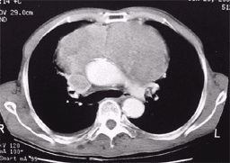
Figure 3 CT Thorax with contrast
Figure 1
Figure 2
Figure 3
Diagnosis:
Thymic carcinoid with superior vena cava obstruction and right sided pleural effusion.
Discussion:
Figure 1 Chest Radiograph
Widening of mediastinum with well defined aortic knuckle - anterior mediastinal mass.
Figure 2 CT thorax (noncontrast)
Huge non-enhancing soft tissue density mass lesion in anterior mediastinum with posterior displacement of the aortic arch and superior vena cava obstruction. Right sided pleural effusion.
Figure 3 CT Thorax with contrast
Huge non-enhancing soft tissue density mass lesion in anterior mediastinum with posterior displacement of the aortic arch and superior vena cava obstruction. Right sided pleural effusion.


