Answer of August 2002
Clinical History:
M/19 Involuntary limbs movement.
Figure 1: Axial Noncontrast CT Brain
Figure 2: Axial T1 weighed MR Brain
Figure 3: Axial T2 Weighed MR Brain

Figure 1
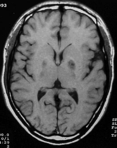
Figure 2
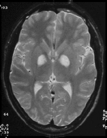
Figure 3
Diagnosis:
Wilson's disease.Putaminal lesions are associated with bradykinesia and dystonia.
Discussion:
Figure 1: There are hypodensities in lentiform nuclei (putamen) bilaterally.
Figure 2 and Figure 3: There are T1 hypointense and T2 hyperintense signal in the putamen bilaterally