Answer of September 2002
Clinical History:
M/35. Fever and cough.
Figure 1: HRCT thorax upper zones.
Figure 2: HRCT thorax at hila.
Figure 3: HRCT thorax at lower zones.
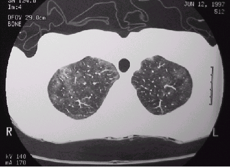
Figure 1
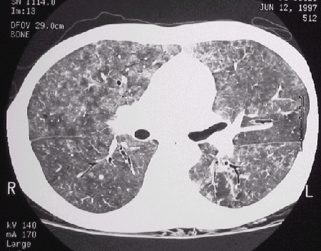
Figure 2
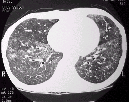
Figure 3
Diagnosis:
Pneumocystis carinii
Discussion:
Bilateral patchy ground glass opacities with areas of normal intervening lung predominately in upper and perihilar regions associated with bronchiectasis and septal thickening. Patient was found to be HIV positive