Answer of February 2003
Clinical History:
M/22. Rhinorrhea.
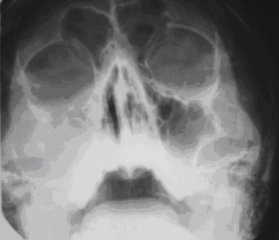
Figure 1
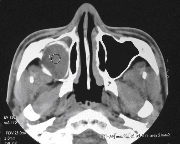
Figure 2
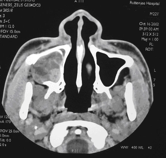
Figure 3
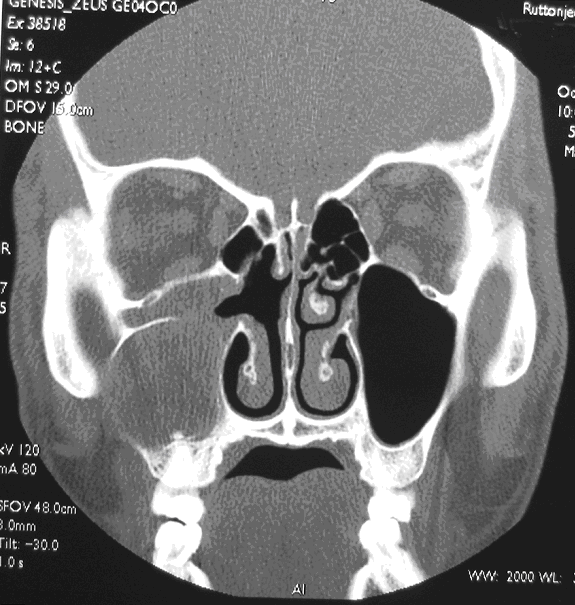
Figure 4
Diagnosis:Discussion:
Ameloblastoma
Figure 1: OM view shows a opacity in right maxillary sinus with bulging of lateral wall of right maxillary sinus.
Figure 2: Noncontrast axial scan of paranasal sinus shows an unilocular lesion with cortical expansion arising from alveolar process of maxilla. It extends superiorly into right maxillary sinus. It extends posteriorly with flattening of fat plane in right retromaxillary space. The lateral wall of right maxillary sinus is deficient with the mass bulging into right buccal space.
Figure 3: The mass shows peripheral enhancement after contrast.
Figure 4: Coronal view with bone window shows involvement of alveolar process of right maxilla.