Answer of March 2003
Clinical History:
F/75, progressive lower limb weakness.
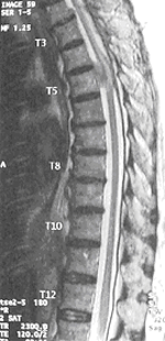
Figure 1. T1 weighed image.
Figure 2. T2 weighed image.
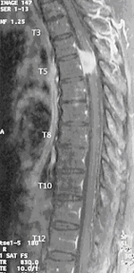
Figure 3. T1 weighed postcontrast image.
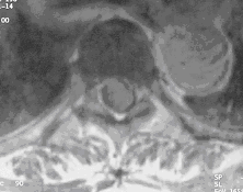
Figure 4. T1 weighed postcontrast axial image at T5 level.
Diagnosis:
Meningioma
Figure 1: T1 weighted sagittal image shows an isointense lesion expanding the cord at T5.
Figure 2: T2 weighted sagittal image shows that the lesion at T5 is isointense.
Figure 3: T1 weighted postcontraast image shows that the lesion is intensely enhanced and appear hyperintense. There is "dural tail" sign.
Figure 4: T1 weighted postcontrst axial image at T5 level suggests that the lesion is intradural extramedullary in location.


