Answer of August 2003
Clinical History:
Abdominal pain. Weight loss.
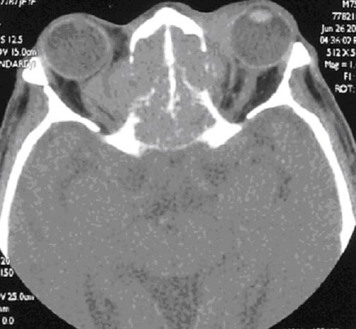
Figure 1. Noncontrast CT abdomen
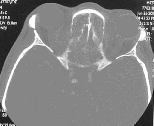
Figure 2. Poscontrast CT abdomen
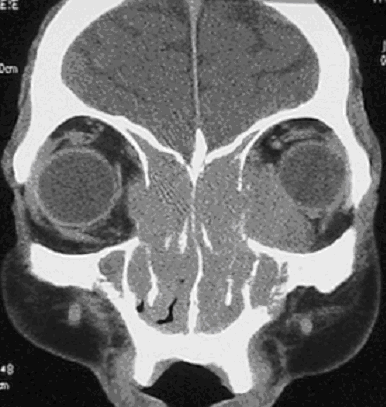
Figure 3. Poscontrast CT abdomen
Diagnosis:
Mesenteric Panniculitis
Discussion:
CT abdomen shows well-circumscribed inhomogeneous fatty mass with higher attenuation than normal retroperitoneal fat extending from mesenteric root toward left abdomen and surrounding mesenteric vessels without distortion. Scattered, discrete nodules of soft tissue density engulfed by hypodense fatty halo are seen inside the mass. It is chronic nonspecific inflammation involving the adipose tissue of the bowel mesentery.


