Answer of October 2003
Clinical History:
M/38 non-smoker. History of chronic rheumatic heart disease and mitral regurgitation. Incidental finding of a solitary pulmonary nodule. Physical examination was unremarkable.
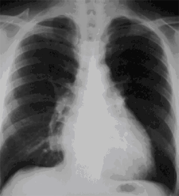
Figure 1: Frontal CXR
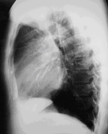
Figure 2: Left lateral CXR
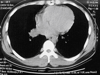
Figure 3: Pre-contrast CT thorax
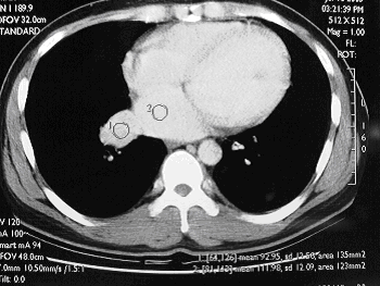
Figure 4: Post-contrast CT thorax
Diagnosis:Discussion:
Pulmonary varix t
- A 3 cm well-defined right paracardiac opacity is seen on the frontal CXR. The opacity is situated in the right lower lobe, as it is posterior to the major fissure on the lateral view.
- The lesion is demonstrated as a dilated right inferior pulmonary vein, draining into the left atrium. It shows comparable contrast enhancement to the left atrium.