Answer of December 2003
Clinical History:
Cough and fever. F/33.
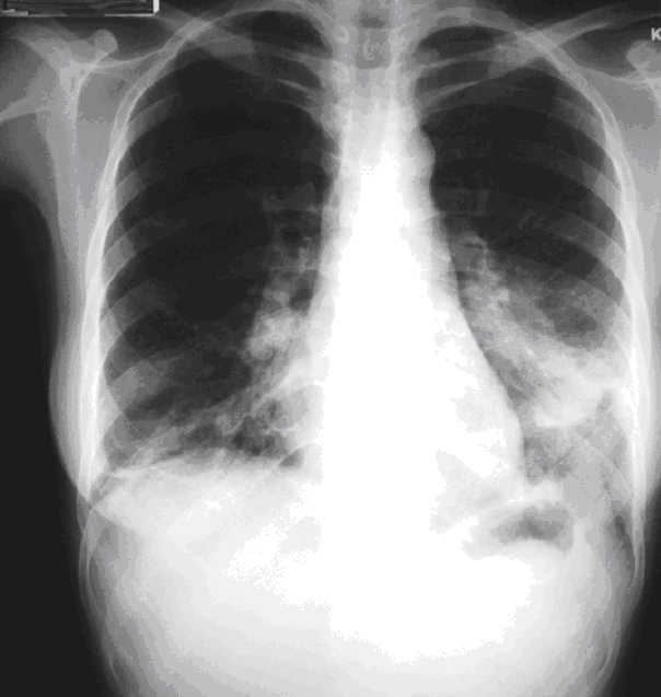
CXR.
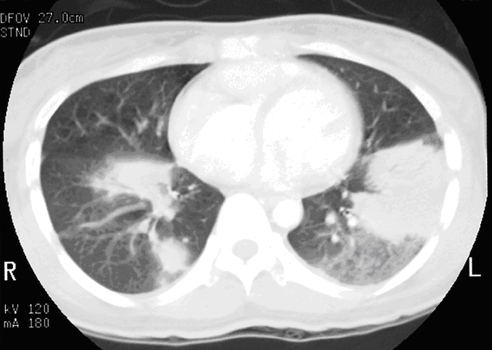
CT Thorax
Diagnosis:
Bronchiolitis Obliterans Organizing Pneumonia
Discussion:
Chest radiographs show multiple nonspecific areas of consolidation in the lower zones of both lungs. CT thorax shows bilateral multifocal consolidation with peribronchovascular distribution. The finding suggests the diagnosis of bronchiolitis obliterans organizing pneumonia. It should be noted that crazy paving pattern is rather a feature of alveolar proteinosis.