Answer of February 2004
Clinical History:
M/38, presented with right scrotal pain.
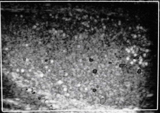
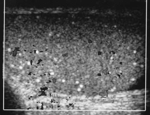
Fig.1 US scan of both testes.
Fig.2 US right testis with colour Doppler
Fig.3 US left testis with colour Doppler
Diagnosis:
Testicular microlithiasis
Discussion:
Ultrasound study of the testes demonstrates numerous tiny echogenic foci bilaterally. They are diffusely distributed and are not associated with distal acoustic shadowing. Symmetrical vascular flow is noted on colour Doppler interrogation.
Testicular microlithiasis is believed to be caused by formation of microliths from degenerating cells within the seminiferous tubules. There has been reported association between this condition and testicular cancer although no causative effect is demonstrated. Association with infertility has also been suggested.


