Answer of May 2005
Clinical History:
69-year-old man with diffuse abdominal pain and decreased general condition.
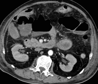
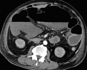
Figure 1. Postcontrast axial CT images of the
upper abdomen in arterial phase.
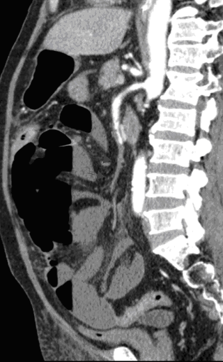
Figure 2. Postcontrast sagittal reformatted CT
image of the abdomen and pelvis in arterial phase.
Diagnosis:
SMA Thrombosis with Bowel Infarct
Discussion:
CT abdomen with contrast in arterial phase showed that there was thrombosis of the distal part of superior mesenteric artery. The associated bowel loops (ascending colon and small bowel) were grossly dilated with no wall enhancement compatible with infarct.
Laparotomy showed gangrenous change of bowel loops from duodenojejunal junction to mid transverse colon.


